
Deeper Questions of Common Sign : Tracking at Kinghurst
This past Saturday was another outing with the Earth Tracks Wildlife Tracking Apprenticeship. We went out to the Kinghurst forest in Grey County, Ontario to see what we could find together. It was a small group of six of us, but that made it a little bit sweeter as we could really dig in to all of the things we were seeing.
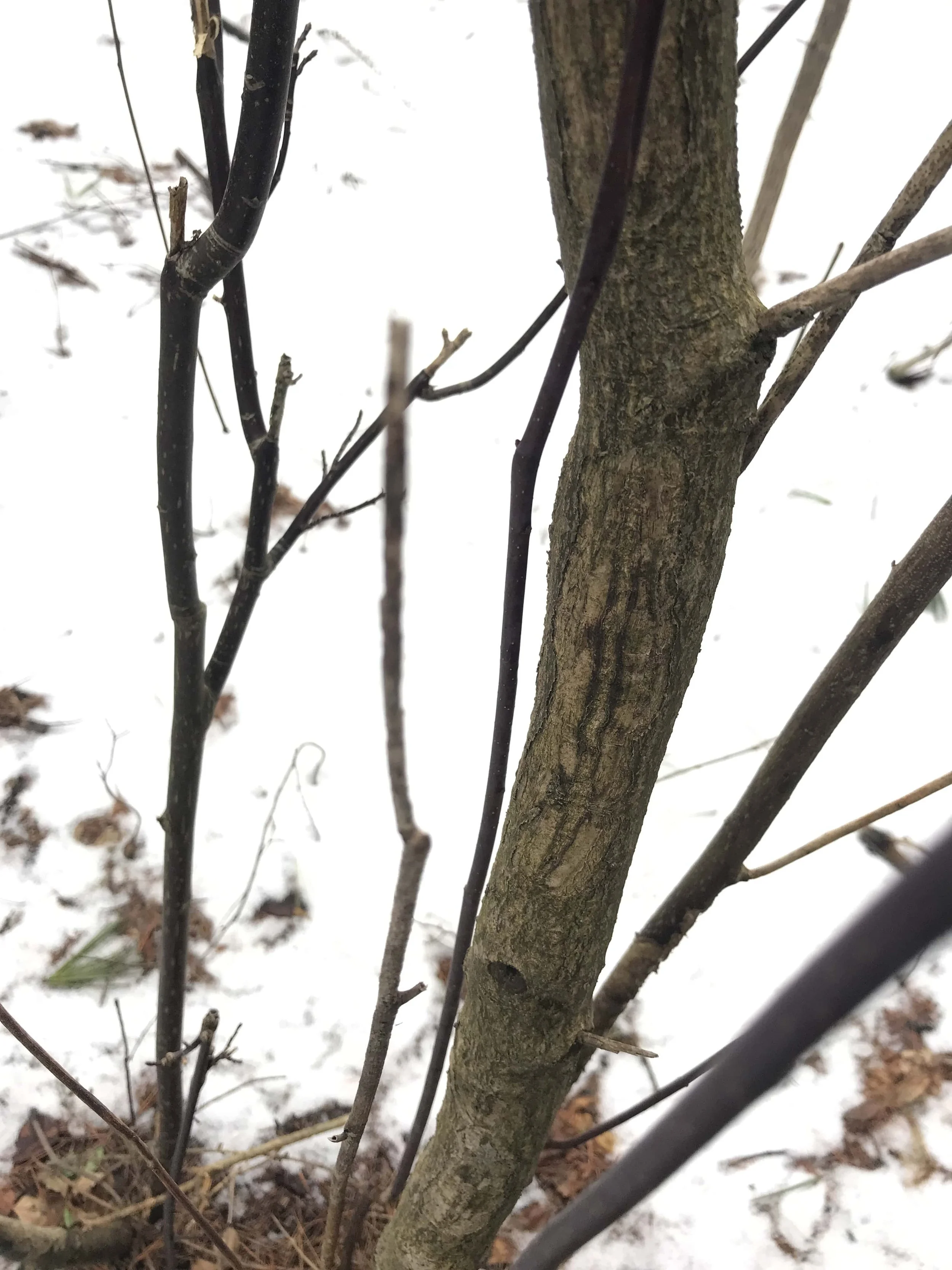
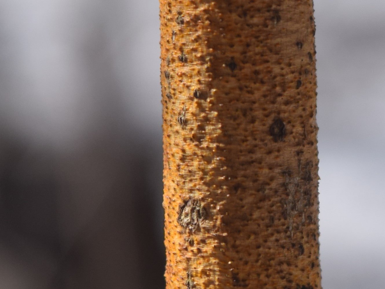
Things got started for me with finding a White Pine (Pinus strobus) tree that had been recently visited by a Pileated Woodpecker (Dryocopus pileatus) who was in search for food deep within the heartwood of the tree.
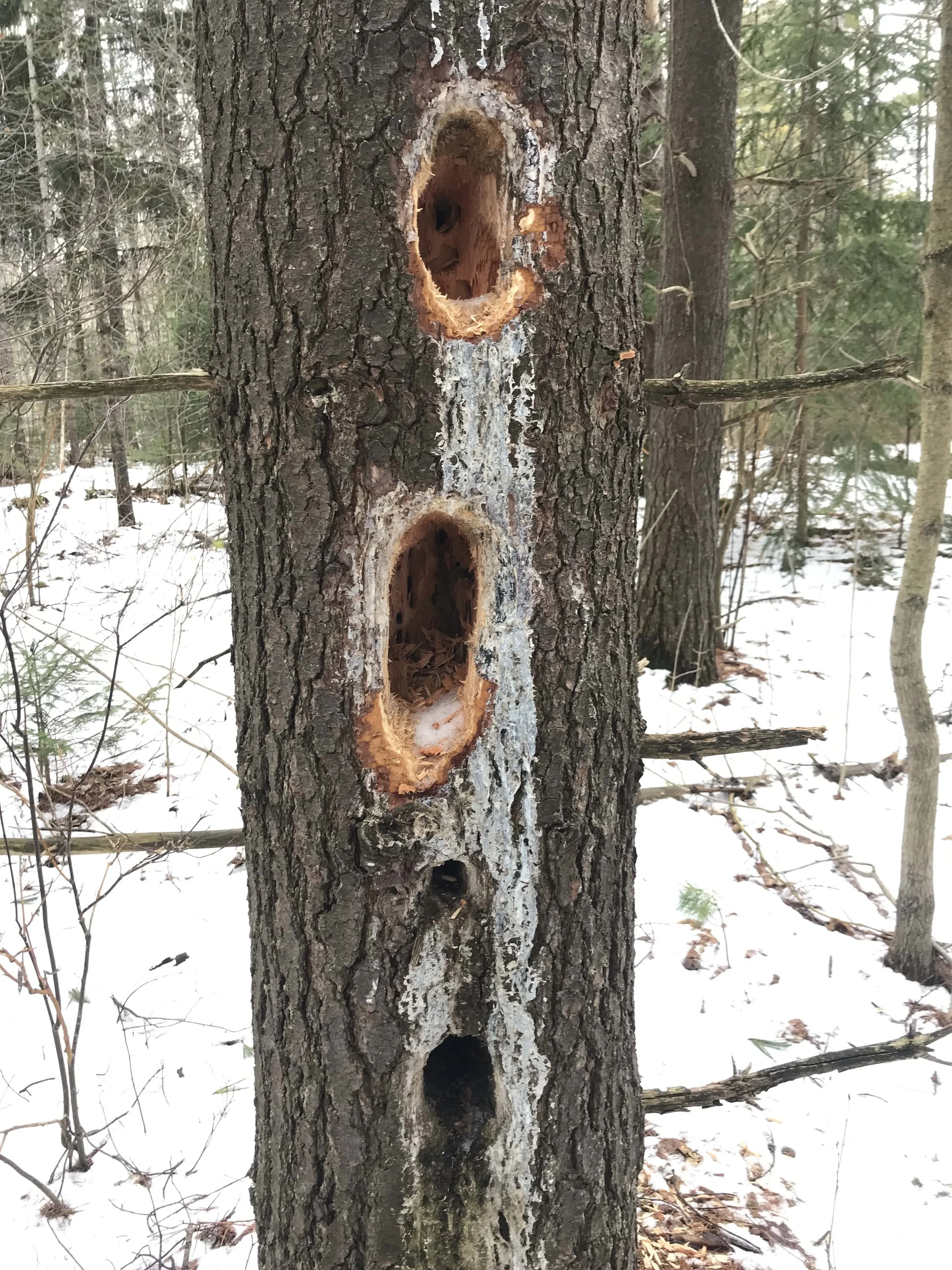
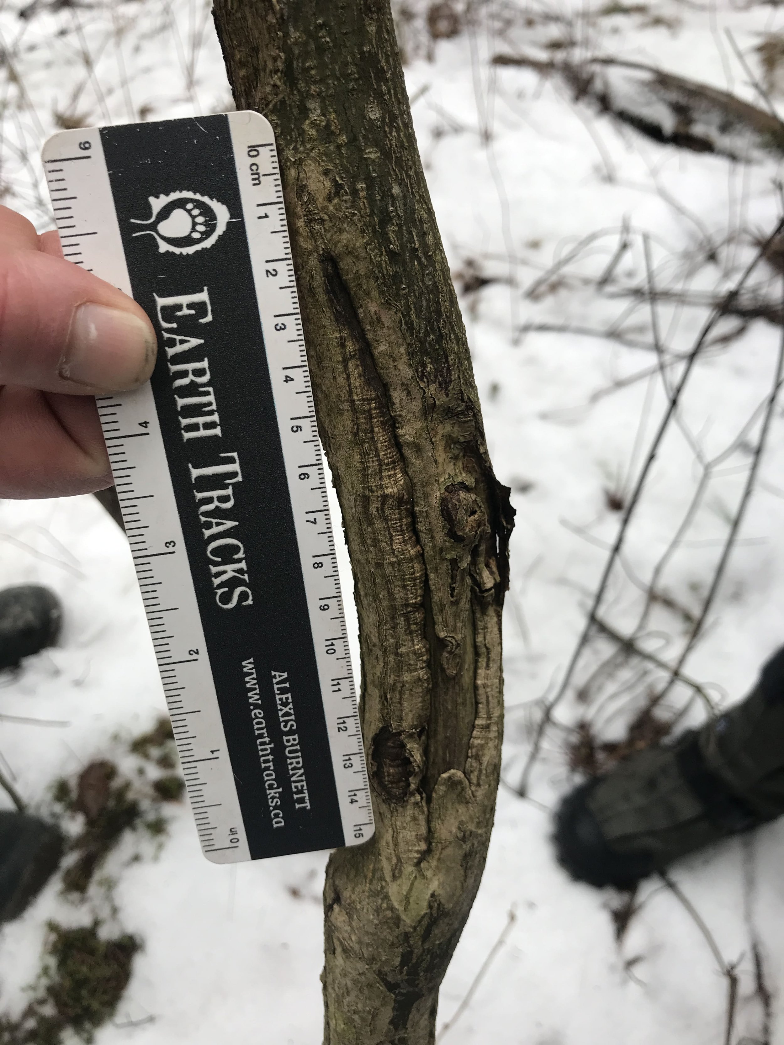
This particular White Pine seemed to have been a regular feeding tree for a while. There were three holes which appeared to be from this Winter, and two lower holes which appeared to be from a previous winter, as the wood inside was dark with debris, a bit of what looked like a speckling mould. While the holes created by the woodpecker were pretty severe, the tree was still quite alive as noted by lot of resinous pitch trying to help scab over the wounds in the tree.
When taking a look onto the ground below I noticed a pile of fairly fresh chips which had been chiseled out from the tree. Within this pile there were a couple of small, somewhat tubular, black and white masses, some covered by the fallen woodchips, some sitting atop of them. I got excited to see these as they are incredible signs left behind by the Pileated, detailing the story of what had occured.
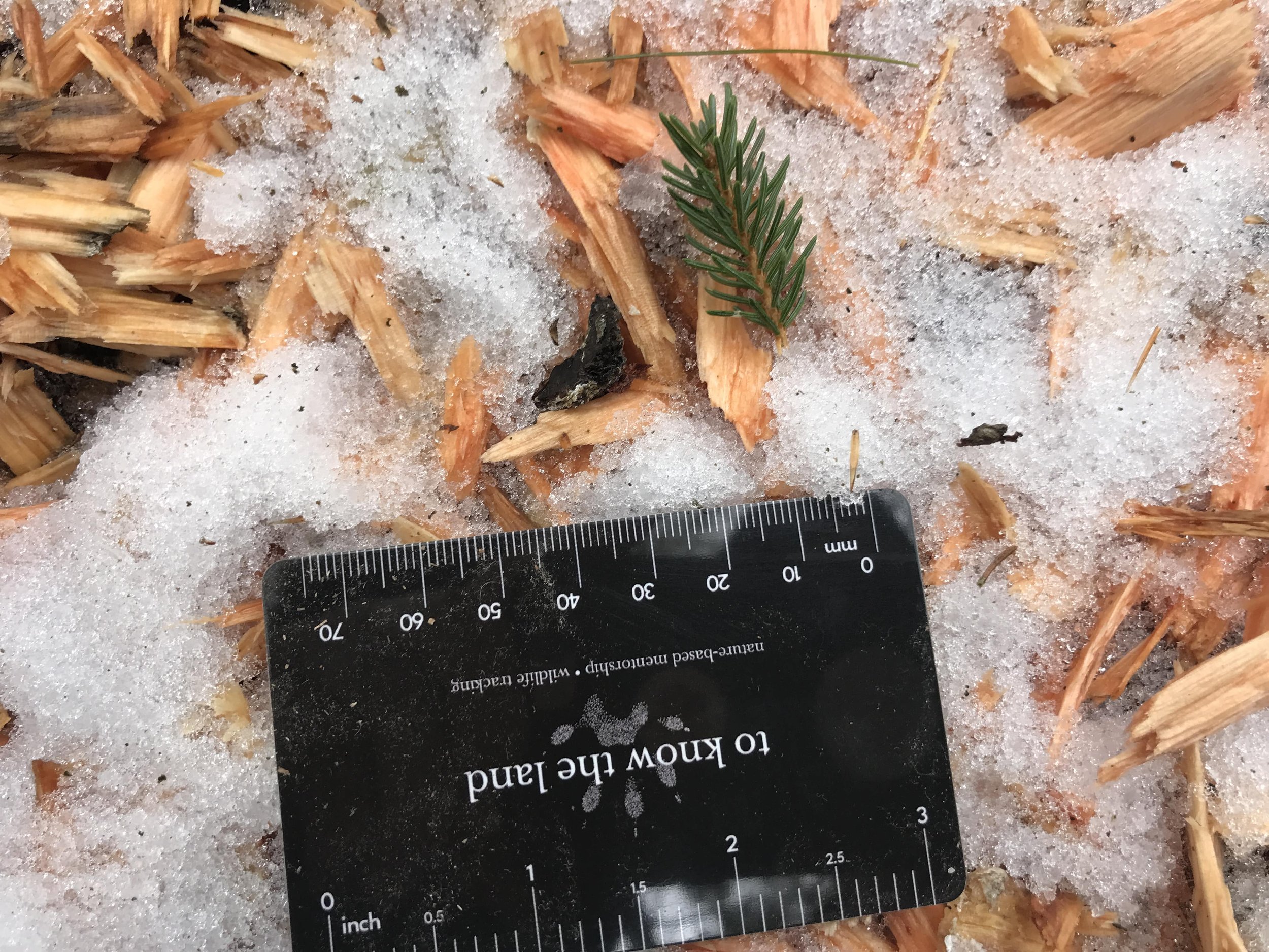
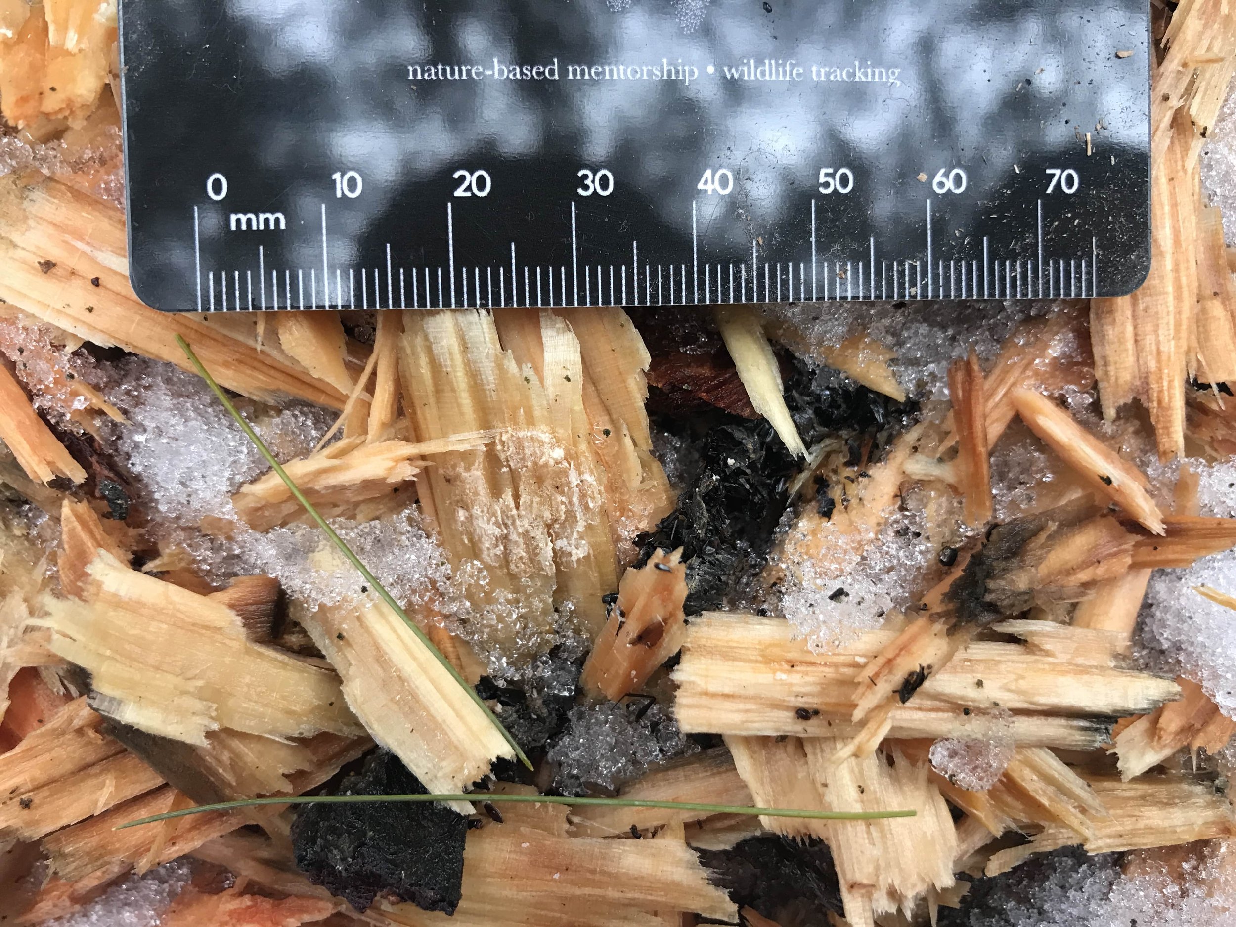
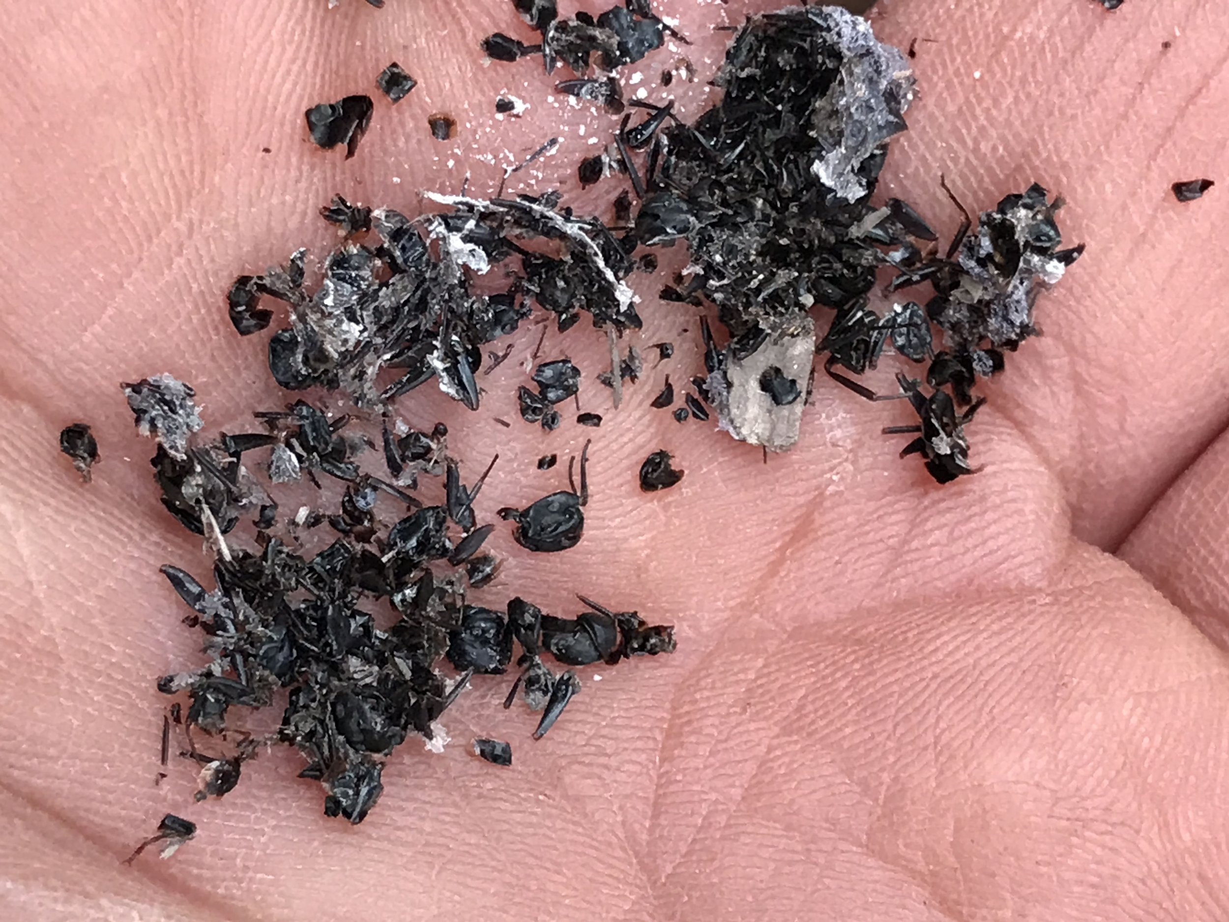
This was Pileated Woodpecker scat, which was made up almost entirely from the chitinous exoskeletons of the Carpenter Ants (Camponotus sp.) the woodpecker had been feeding on from the excavated holes. The Carpenter Ants were in the heartwood where they too excavate the wood, creating colonies where they lay eggs, rear their young, and live out their lives. I have read in Tracking and the Art of Seeing by Paul Rezendes that the Pileated listens for the ants in the tree. This could make sense in the Summer when the Carpenter Ants are active, but in the Winter, when the ants are in diapause, how do the Pileateds find the ants? Are they just returning to trees where they know the ants have been in the past? Are they using other senses to find the ants? What about smell? The smell of the tree resins would be pretty powerful and may cover up the scent of any formic acids the ants are producing in the colony, and I actually don’t know if Pileateds have a good sense of smell. Perhaps they don’t?
Another thing I was noticing about this tree is that the older holes are lower on the trunk of the tree and the newer holes are higher up. I have heard and seen that the behaviour of Pileated Woodpeckers excavating lower in the tree is done in Winter as the Carpenter Ants migrate to lower areas on the tree as the ground temperature is warmer than the ambient air temperatures higher up. If this is true, could it then be true that the Carpenter Ants may only go as low as they are compelled to by the cooling Autumn or Winter temperatures? Could we use these woodpecker excavation holes to determine relative chill of the Winters when they were produced? Was the Winter colder when the older holes lower on the trunk were created?
Another question which may be worth looking into in the future would be how the Pileated Woodpeckers deal with the sticky resins which must get all over their bills and perhaps their feathers, too. Do they rub their bills on branches as a bird of prey might when they are cleaning their bills of the gore from their recent meal? Does their saliva help break down the pitch? Do they have saliva? So much to know..
For years I have been identifying a shrub, the Alternate-leaved Dogwood (Cornus alternifolia), otherwise known as Pagoda Dogwood because of the habit of branching in an alternate pattern (leaves branching out from the main stem in a staggered habit) as opposed to most other dogwoods which branch in an opposite pattern (leaves and branches coming off of the main stem in opposite each other, like how our arms branch out from our “trunk” from the same point along the length of our body).
We were checking out the Alternate-leaved Dogwood because we noticed some possible old incisor scrapes on the narrow trunk. Alexis pointed out the incisor marks and mentioned that this was a favorite shrub of the White-tailed Deer (Odocoileus virginianus) to feed on in this part of the world. I hadn’t noticed the faded grey wood which appeared exposed for at least a year. I wondered at why they were incisor scrapes instead of antler rubs? If I remember correctly, Alexis pointed out that the width of the scrapes if taken individually would be too narrow for an antler rub. Also, the height where they were on the shrub may be a little too high for an antler rub. I believe Renato or perhaps Tamara pointed out the clean lines where the teeth entered into the bark at the base of the scrape, as well as the clean exit holes where the deer pulled the bark off of the trunk would be unlikely in the case of the more abrasive antler rubs, which tend to leave frayed uneven edges at the tops and bottom of the wounds in the bark.
In Winter, sometimes a faster way to identify the Alternate-leaved Dogwood is by noticing that sometimes the branches can be a golden yellow colour which appears bright amidst the drab of greys, browns and white. We noticed the same thing while we were out tracking. Sure it wasn’t tracking an animal, but noticing how plants interact with their environments and who interacts with the plants can still teach us about a place, and that is really my end goal. I had noticed this many times before in my neck of the woods and I was able to share that this wasn’t necessarily an identifying feature of the dogwood, but instead a field mark of an entirely different species which was infecting the dogwood!
Especially seen in Winter when the Pagoda Dogwood has lost all their leaves, the Golden Canker (Cryptodiaporthe corni) slowly infects and kills twigs, small branches, and possibly even the main stem of the shrub. Golden Canker is an Ascomycete fungi which likely infects the shrub though wounds in the bark, lenticels (pores in the bark for gas exchange), leaf scars (where leaves were once attached to the twig), or broken limbs though this remains uncertain. When I often see the canker infecting Pagodas along the sides of trails, I have wondered if these particular shrubs were infected through wounds acquired through broken limbs from cyclists or folks running by and breaking off branches accidentally? The fungus gets under the protective bark and may just chill in the tree for a while, not really causing any problems. But something, perhaps drought, or too much Sun, causes stress to the shrub, then the canker can start to reveal itself and the shrub begins to show signs of infection.
The canker appears as small yellow to orange spots on the tree which are the fruiting bodies of the fungi. In the Spring the fruiting bodies release spores which can infect other nearby Pagodas or even other parts of the same plant. In warmer months the mycelium beneath the bark spreads and the canker can infect more of the branch or stem it is growing on. Eventually, as the canker spreads and kills off living tissues of the tree, the branches the fungi is growing on will no longer produce leaves, and soon will die. Sometimes the shrub can isolate the infected branch by compartmentalization, where the shrub will allow the branch to die off at a node before the mycelial hyphae can spread beyond the node, thus protecting the main stem. Sometimes this works, sometimes not. I have read that sometimes the fungi spreads and infects the main stem and can kill the shrub, but I have yet to see this myself, but I’ll be looking now.
I remember when I first looked up Golden Canker; it wasn’t because I wanted to know which fungus was killing off the branches, I didn’t even know it was a fungus! It was from a pure sense of wonder, a question: Why were some of the branches of the Alternate-leaved Dogwoods I was finding yellow? By framing the curiosity in a question I changed how I engaged with it. I was then compelled to look it up, to investigate the unknown. If I tried to label it right away, I would have come to some conclusion and been stuck in a, likely incorrect, diagnosis and never really learned about the relationship between the canker and the shrub. By leaving the question open ended it allowed for all sorts of possibilities, none more possible than another. If I had framed the question in a closed way, with a simple answer, I may not have investigated further. All this reminds me that questions can be more valuable sometimes in trying to see and learn new things than unfounded conclusions and answers are. It reminds me to keep a sense of wonder if I want to learn to see which I cannot yet see in the world.
We kept on, finding what appeared to by either Coyote (Canis latrans) and/or Domestic Dog (Canis familiaris) tracks and trails. We followed them for a while, measuring strides and individual tracks of the feet trying to find any discriminating sign, but I personally, I couldn’t really settle on one or the other, though the trail did seem rather straight, and didn’t waver much, though the animal may have turned back towards the direction of the parking lot, a behaviour which I usually associate with a dog returning to their human (Homo sapiens).
We also began looking at different squirrel trails, including the Red Squirrel (Tamiasciurus hudsonicus) and Grey Squirrel (Sciurus carolinensis). I have heard of different ways of telling them apart but my go to is the trail width. Measurements collected from Mark Elbroch’s Mammal Tracks and Sign indicate that a Red Squirrel trail width for a bounding gait is 7.3 - 11.1 cm (2⅞ - 4⅜ in), while a Grey Squirrel trail width in a bounding gait is 9.5 - 15.2 cm (3¾ - 6 in). This shows an overlap and so I wonder, what are some other distinguishing features to look for in a blurry trail?
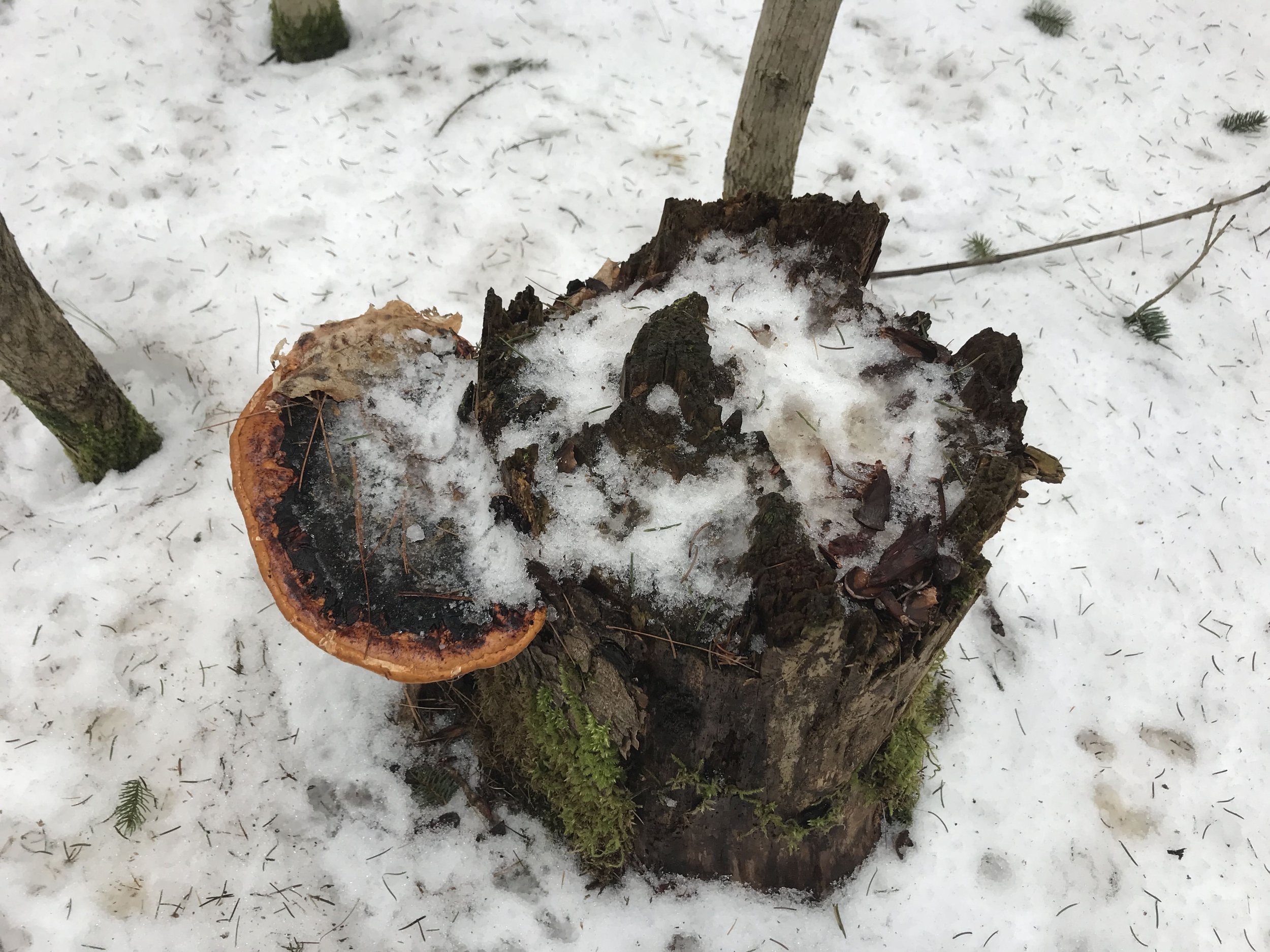
What about stride length? High end of a bounding stride of a Red is 63.5 cm (25 in) and a Grey 76.2 cm (30 in), which is 12.7 cm (~5 in) which isn’t much to go on with. Anything else? Sometimes following out a trail can tell us a lot. So that is what I did a couple of times. First one led to a White Pine (Pinus strobus) with a Red Squirrel watching me from the base of the trunk, which was a bit of an easy giveaway. The other led to a ran along a log, past a stump with a Red-belted Polypore (Fomitopsis pinicola) all the way up to a midden pile of old Black Walnut (Juglans nigra) shells. The shells were chewed up on both wider sides and all but a couple with what appeared to be incisor scrapes along the midrib. The few without had no real midrib remaining. This is pretty tell-tale signs of Red Squirrels.
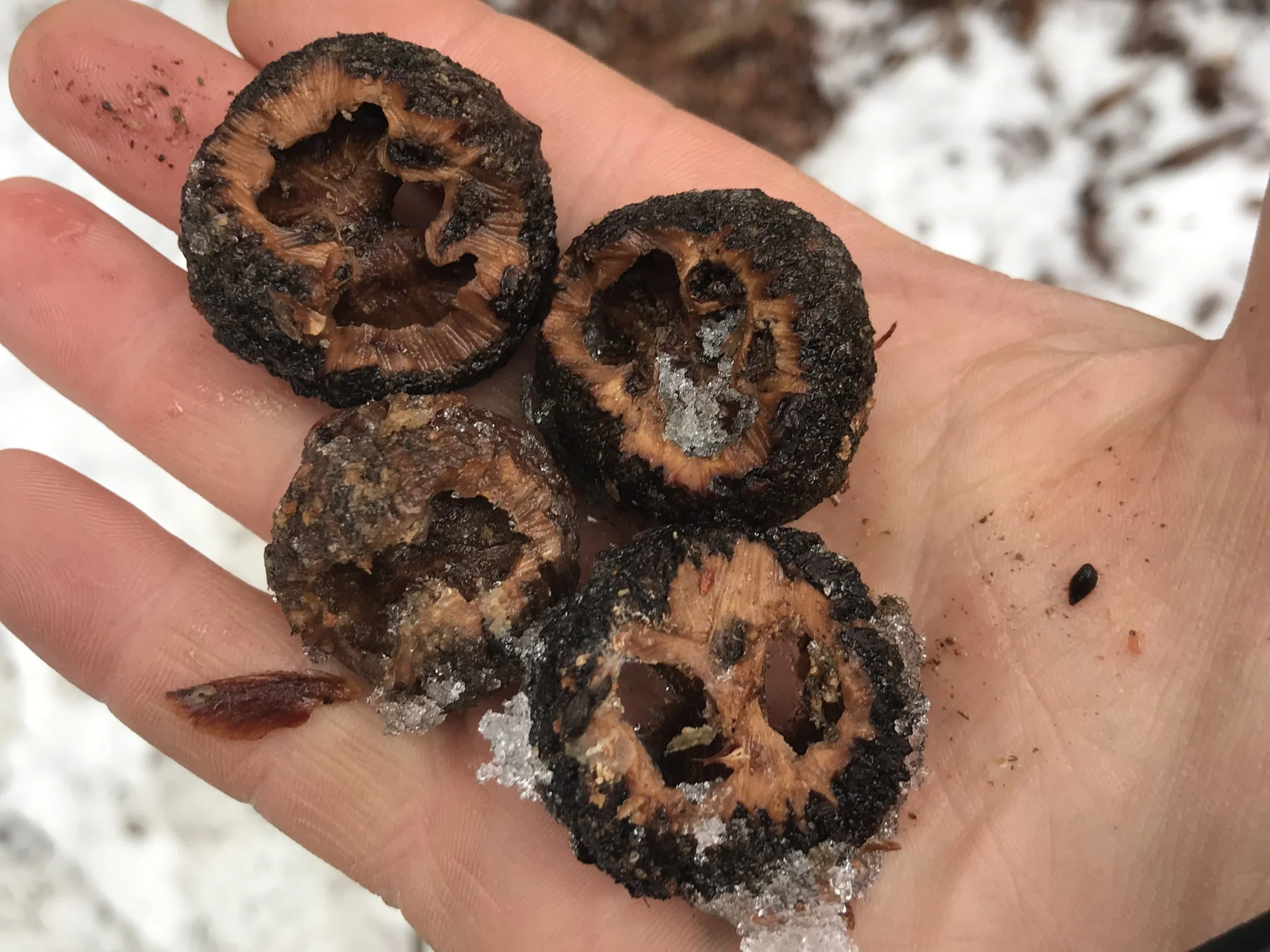
Elbroch writes for Grey Squirrels “[l]ook for their signs spread below many perches, rather than accumulating in large piles below just several perches…” which would be more common for the Reds. Seems like a Red to me.
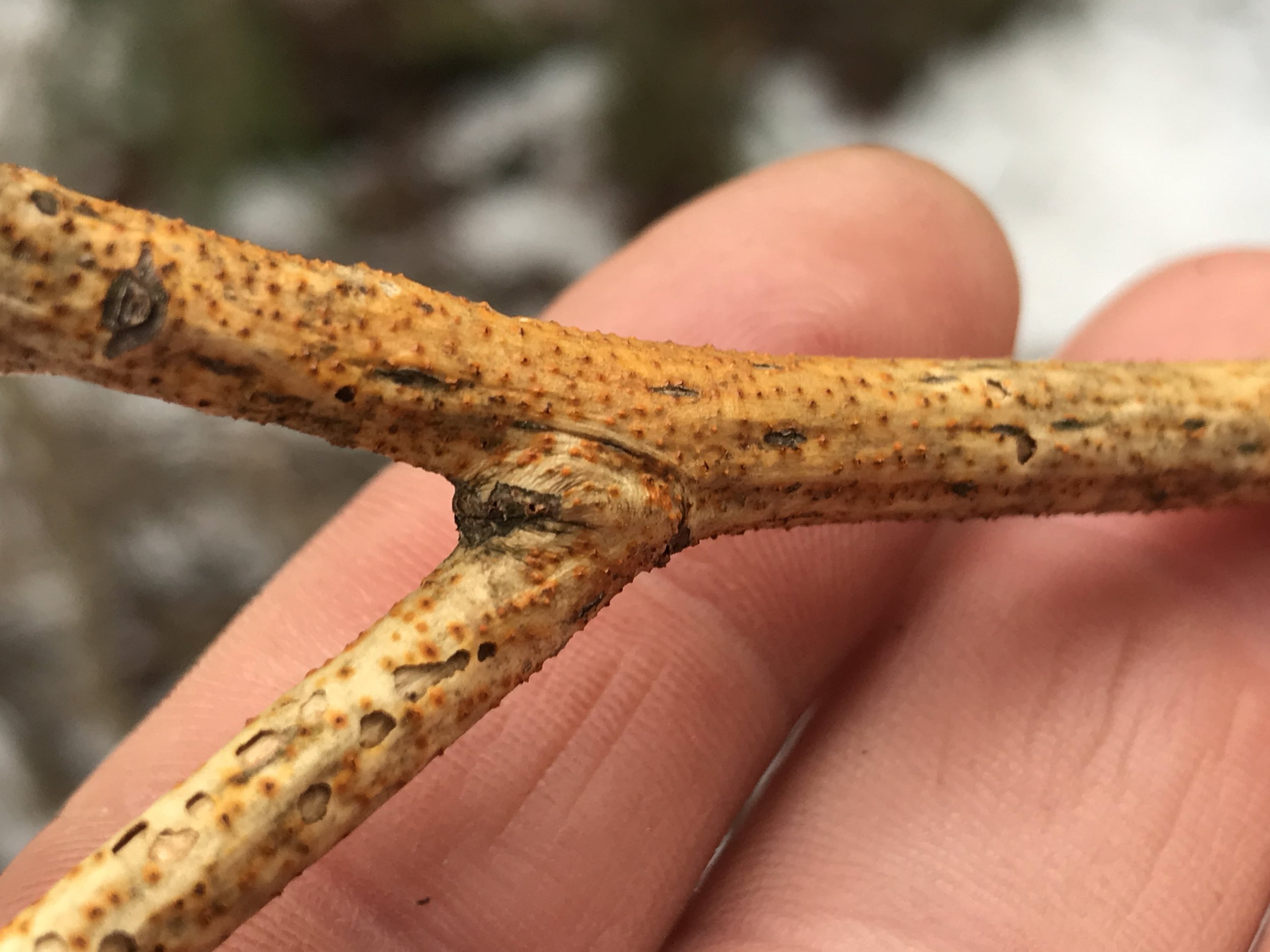
It wasn’t until a moment later when Alexis asked if I had seen the incisor marks on the polypore? I responded that I did not, but I quickly returned to investigate.
Red-belted Polypores are often found growing on trees in the Pine family, hence the specific epithet “pinicola”, and they have been used in medicine for immune support due to their anticancer, antifungal, and antibacterial qualities (Hobbs, 2020). As I looked at the bracket fungi growing on the stump I wondered if the animal who had consumed some of the outer ring, the newer growth, had understood these properties and was consuming the fungi for these reasons?
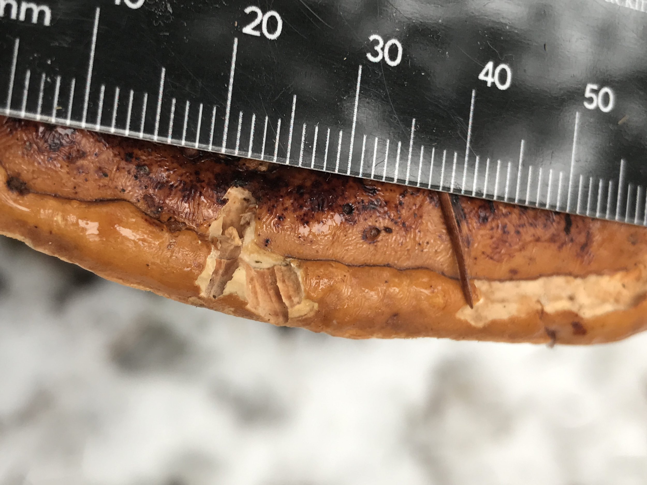
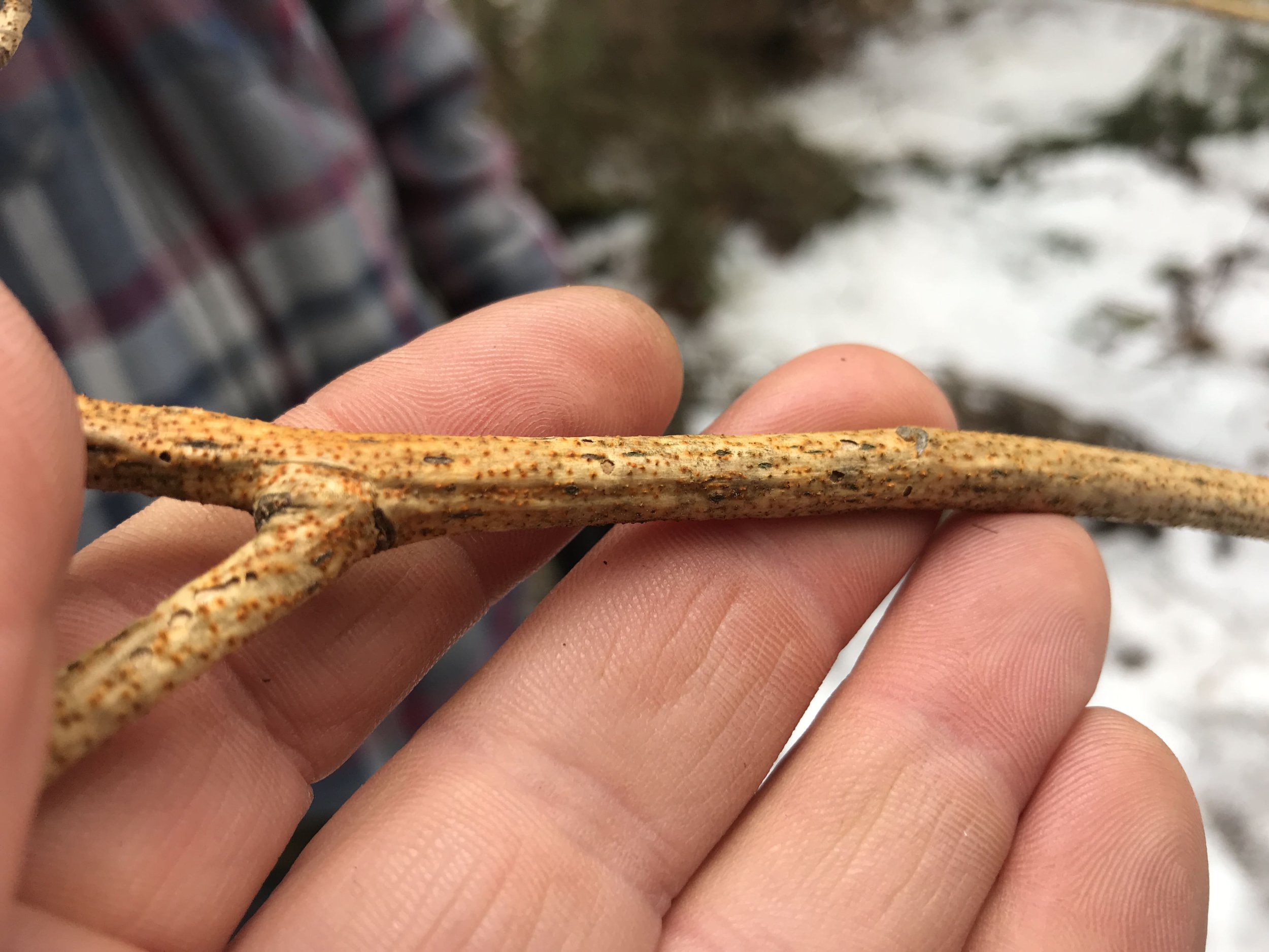
My first thought when looking at the fungi was that the Red Squirrel had been feeding on it, but on closer inspection, I started to have my doubts.
Often rodents anchor into a mushroom with their lower incisors and then cut into the mushroom with their upper incisors, cutting away a morsel to eat as they bring the uppers down to meet the lower. Imagine biting into an apple. Now when this happens on a tougher shelf mushroom like the Red-belted Polypore sometimes the incisor marks stick around. The issue I took with the Red Squirrel possibility was the width of the cuts of each of the incisors. They just seemed too small for a Red Squirrel.
According to Mark Elbroch’s book Animal Skulls (Stackpole Books, 2006) Red Squirrel incisors measure 1.06 mm at the smallest. This is even a measurement of the bottom incisors which I do not believe to be the ones used in cutting away a bite of mushroom. I just use this number to illustrate that the smallest data point is still too large for the sign left behind. In light of that, we must assume someone else was mowing down on the mushroom. Two other small rodents in the area would be Deer Mouse (Peromyscus maniculatus) and Southern Red-backed Vole (Clethrionomys gapperi) both of whom may consume fungi. Deer Mouse upper incisor measurements range from .64-.86 mm, with an average of .73 mm. For the Southern Red-backed Vole, the uppers range from .65-93 mm, with an average of about 77.5 mm. These numbers are closer to the the width of the incisor cuts which I estimate at around .7-.8 mm. I estimate because my ruler has relatively thick lines in comparison with the incisor scrapes and no delineation between the millimeter markings to determine the exact number (maybe I’ll ask for a good caliper set for my birthday). Both the Deer Mouse and Southern Red-backed Vole have many recorded instances of consuming fungi, though I cannot find a paper or published article about them particularly feeding on Red-belted Polypore. All in all, I cannot name who I believe it to be between the Red-backed or the Deer Mouse, but I do not believe I saw feeding sign from a Red Squirrel.
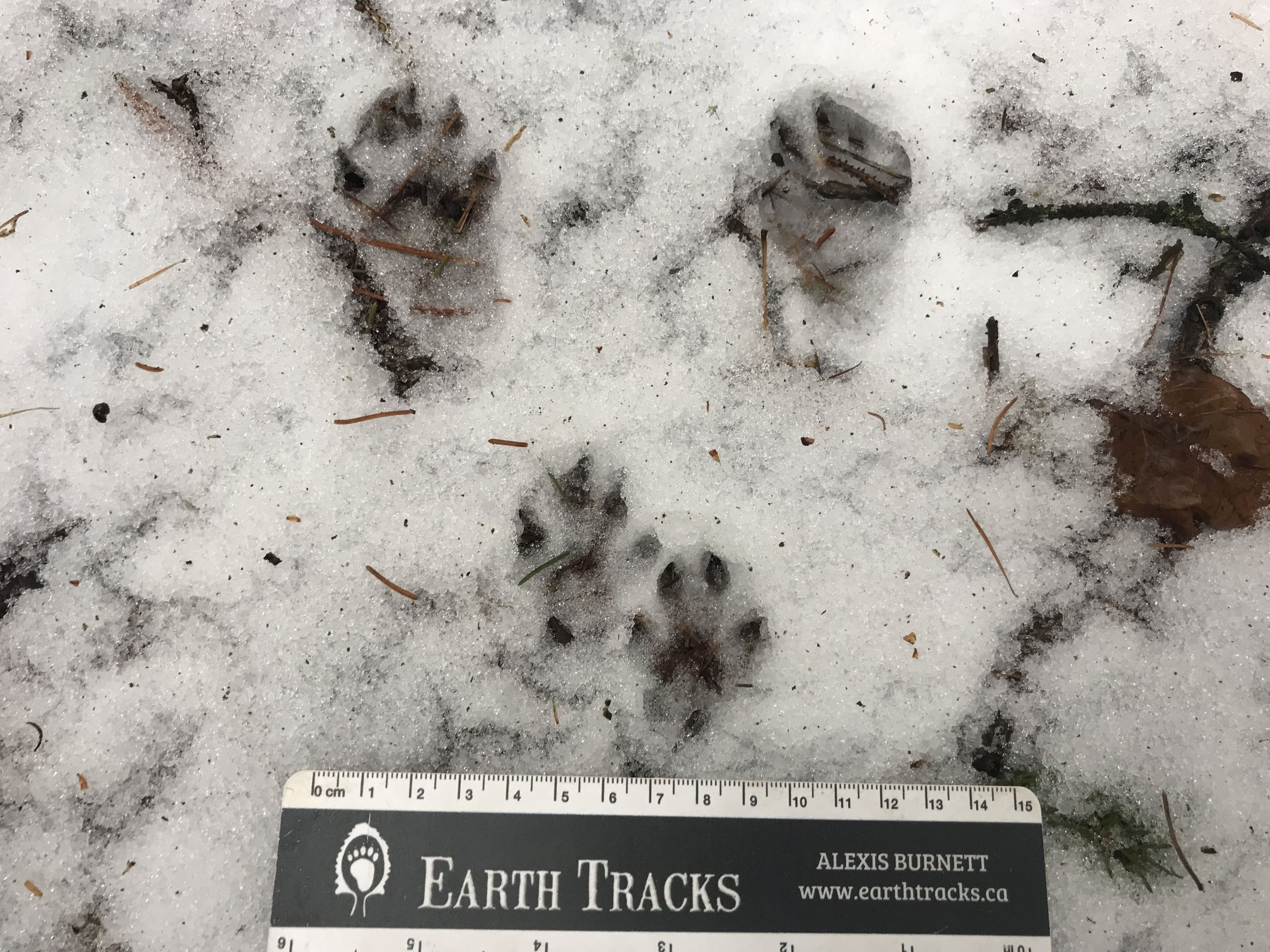
As we went on there were some questions around gaits which I thought were pretty fun and I was curious about looking into further. Here is a photograph of one of the gaits.
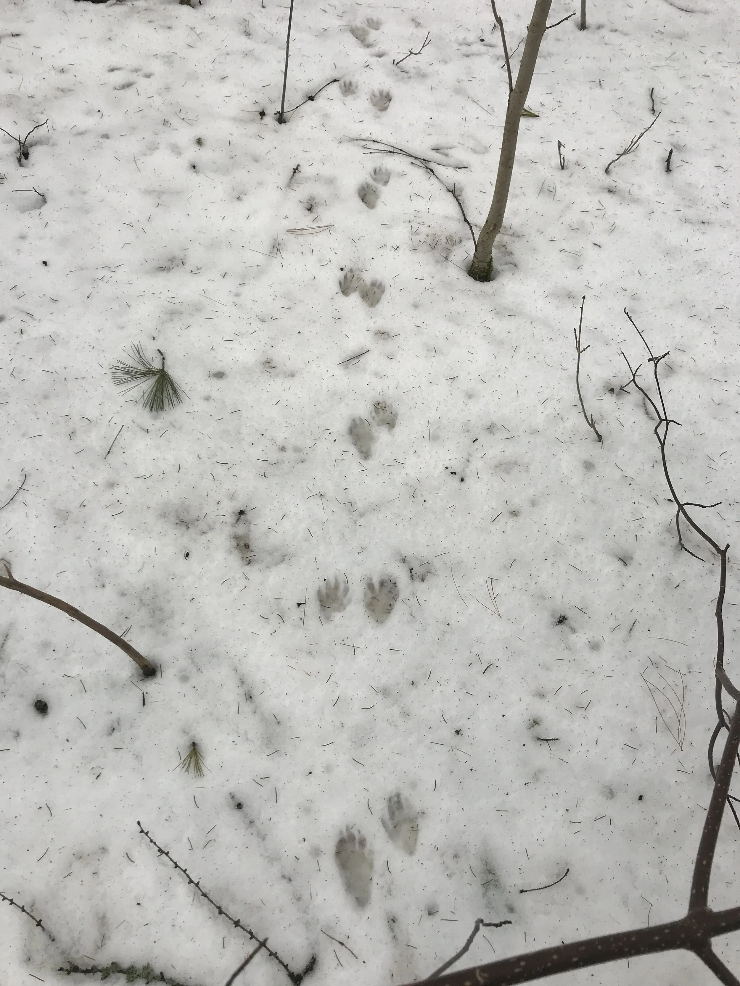
If you look carefully you can see five toes on either foot, with an alternating patter of a longer tracker beside a somewhat shorter one. This is the trail of a Raccoon (Procyon lotor) whose trail leaves behind the track pattern of their fronts and hinds landing beside each other. Everyone in our group agreed with this assessment. We were wondering which gait created this pattern, are there multiple gaits that leave this pattern, and what was each gait properly called? Gaits are called different things by different authors and sometimes between different animals, notably equine gaits. But I’ll do my best to parse out the details below.
Raccoons move in all sorts of gaits, but they, like most animals, have a preferred set of gaits that they commonly use. One of these gaits is called a pace. A pace is described as when the front and rear on the same side of an animal step forward in unison or nearly so. If I was on all fours and my right front and right hind step forward at the same time and then come down at the same time, or nearly so, and then my left front and left hind step forward and land together and this pattern is repeated over and over, than this is a pace. The University of Minnesota College of Veterinary Medicine website on gaits defines a pace as “a two-beat gait. The two lateral limbs are used alternately for weight support, i.e., the left forelimb and left hindlimb move in unison, as do both right limbs. The forelimb may cycle slightly earlier than the ipsilateral hindlimb.” It is a gait shared by Raccoons, Bears (Ursus spp.), Mountain Lions (Puma concolor), as well as Camels (Camelus spp.), Giraffes (Giraffa spp.), and members of the Elephantidae family. The pace is a gait that doesn’t require as much energy as a trot and has a lower arc of stride, or vertical oscillation in the movement. It’s a pretty chill gait for a Raccoon to use. I have also heard this gait called a waddle since the body sways laterally back and forth as the animal moves forward. I believe that a pace is actually the same thing as a 2x2 walk, but is just a different name. If anyone reads this and disagrees, please let me know. I am here to learn!
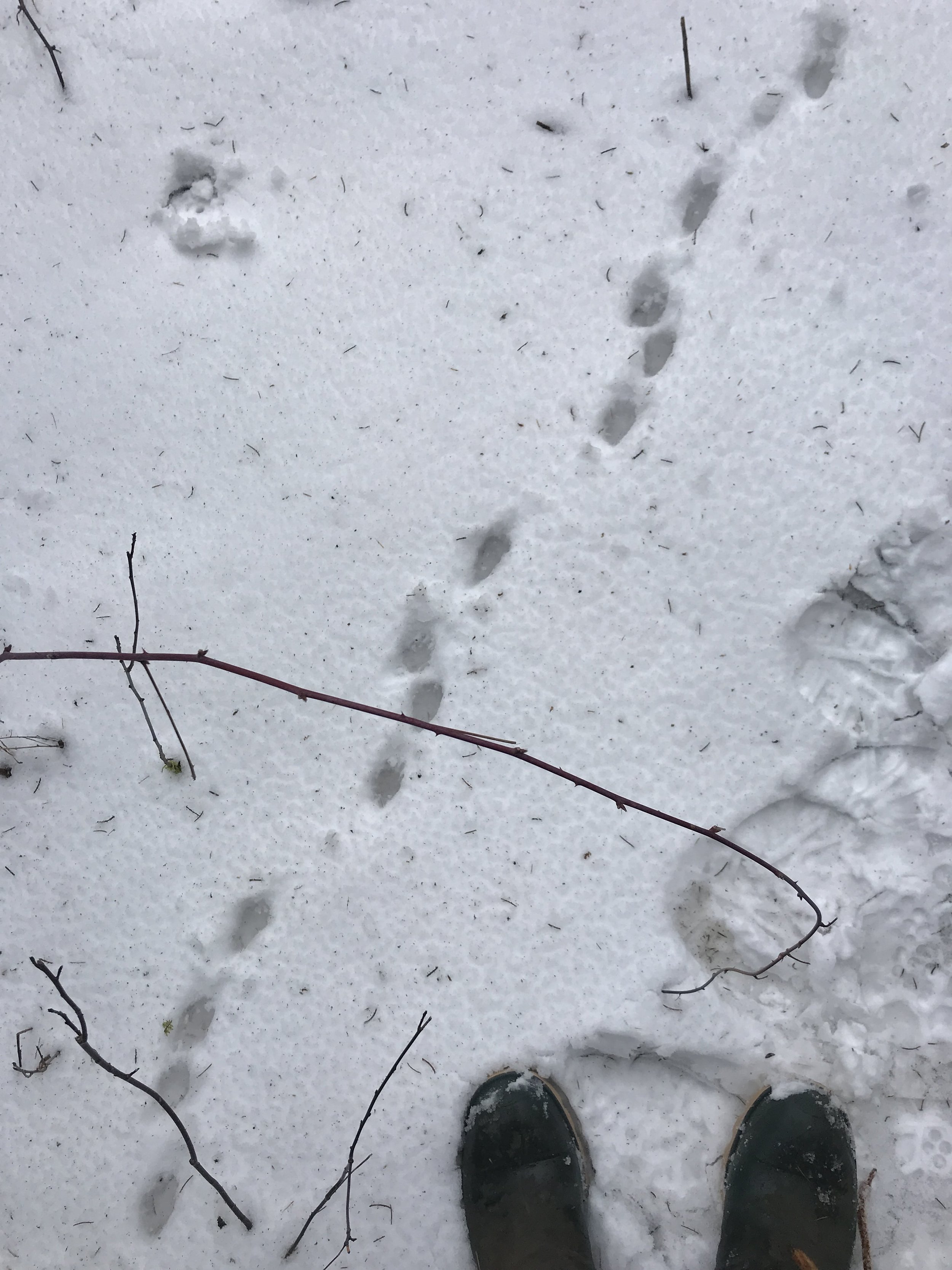
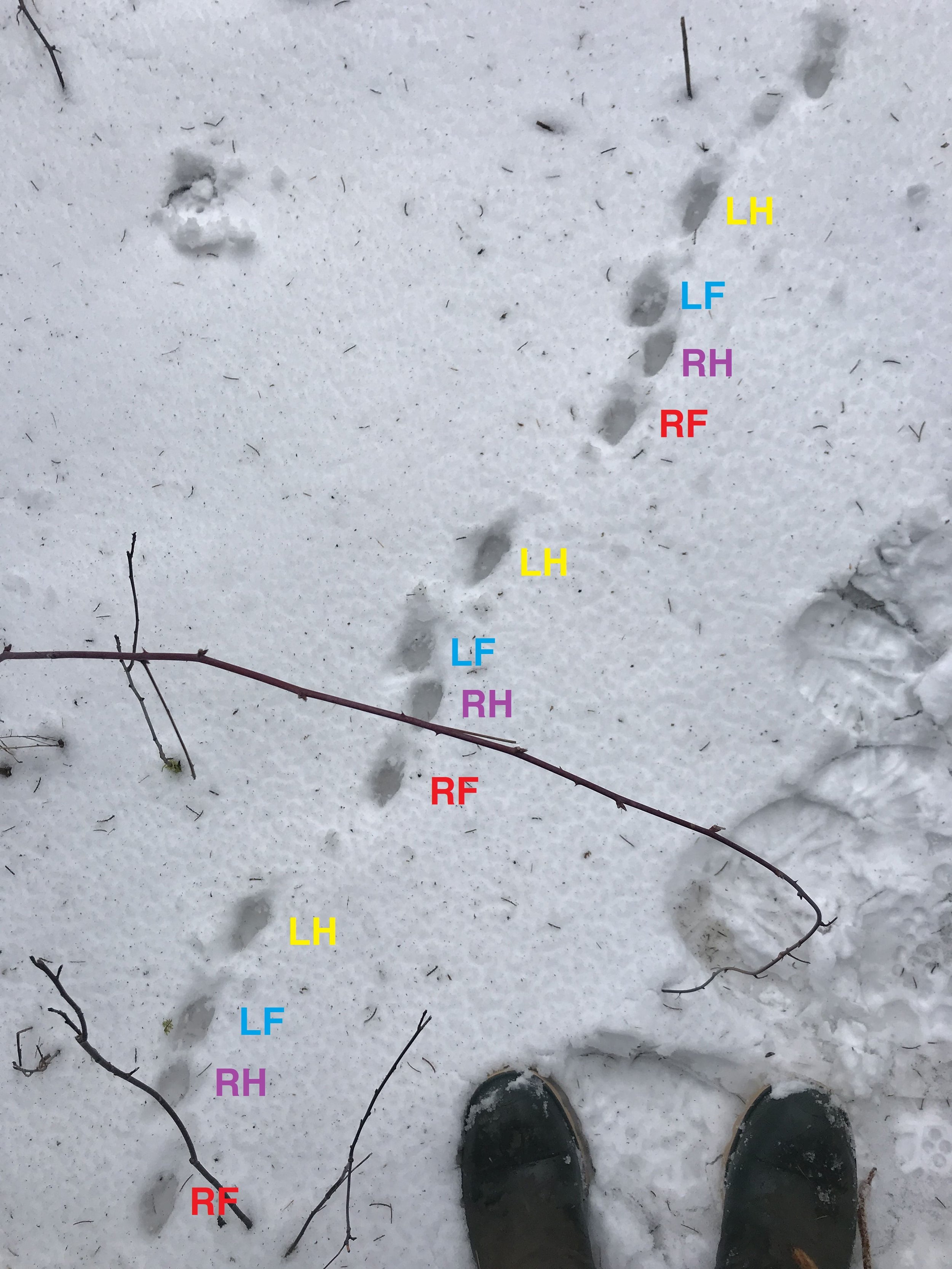
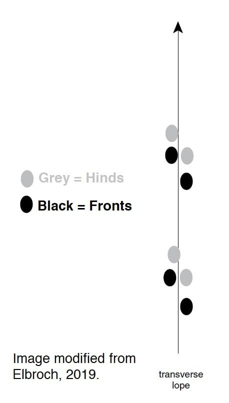
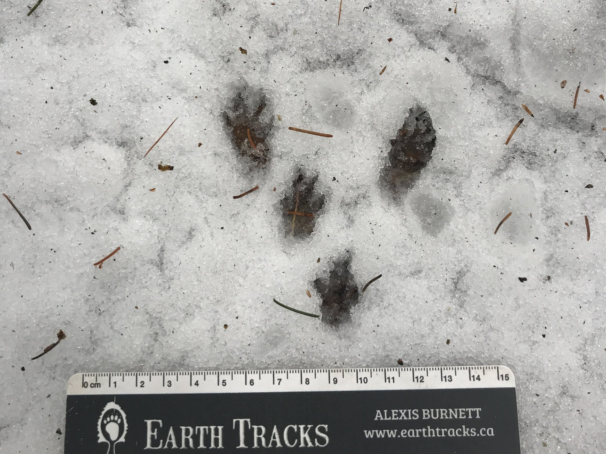
Right before we sat down for lunch under a skirt of Spruce branches (Picea sp.) we came across another trail. This trail changed patterns a couple of times, and considering this frequent change, and based on the more rectangular shape of the tracks, the narrow trail and overall size of the tracks we recognized them as s Striped Skunk (Mephitis mephitis). Commonly I see a track pattern representing a transverse loping gait for the Striped Skunk, but it does seem to switch patterns pretty frequently. Let’s review the gaits in the first photo (the one with the boots). From the bottom left I see a group of four tracks made up of a right front, right hind, left front, left hind. This we would call a transverse lope for two reasons; the hind feet do not entirely pass the fronts making this a lope and not a gallop. Secondly, we call it a transverse lope because the trail shows a pattern off footfall going across the body, with the right front landing first, and then the left front. As the left front is leaving the ground the right hind touches down beside where the left front was previously. After the right hind touches down it is followed by the left hind. Both hinds then push off and create a bit of a gap where the animal is temporarily airborne for a moment before the right front lands again in the next group of four. Note that these groups of four are also indicative of a lope where as an overstep walk would be in groups of two, but we’ll take a look at that below.
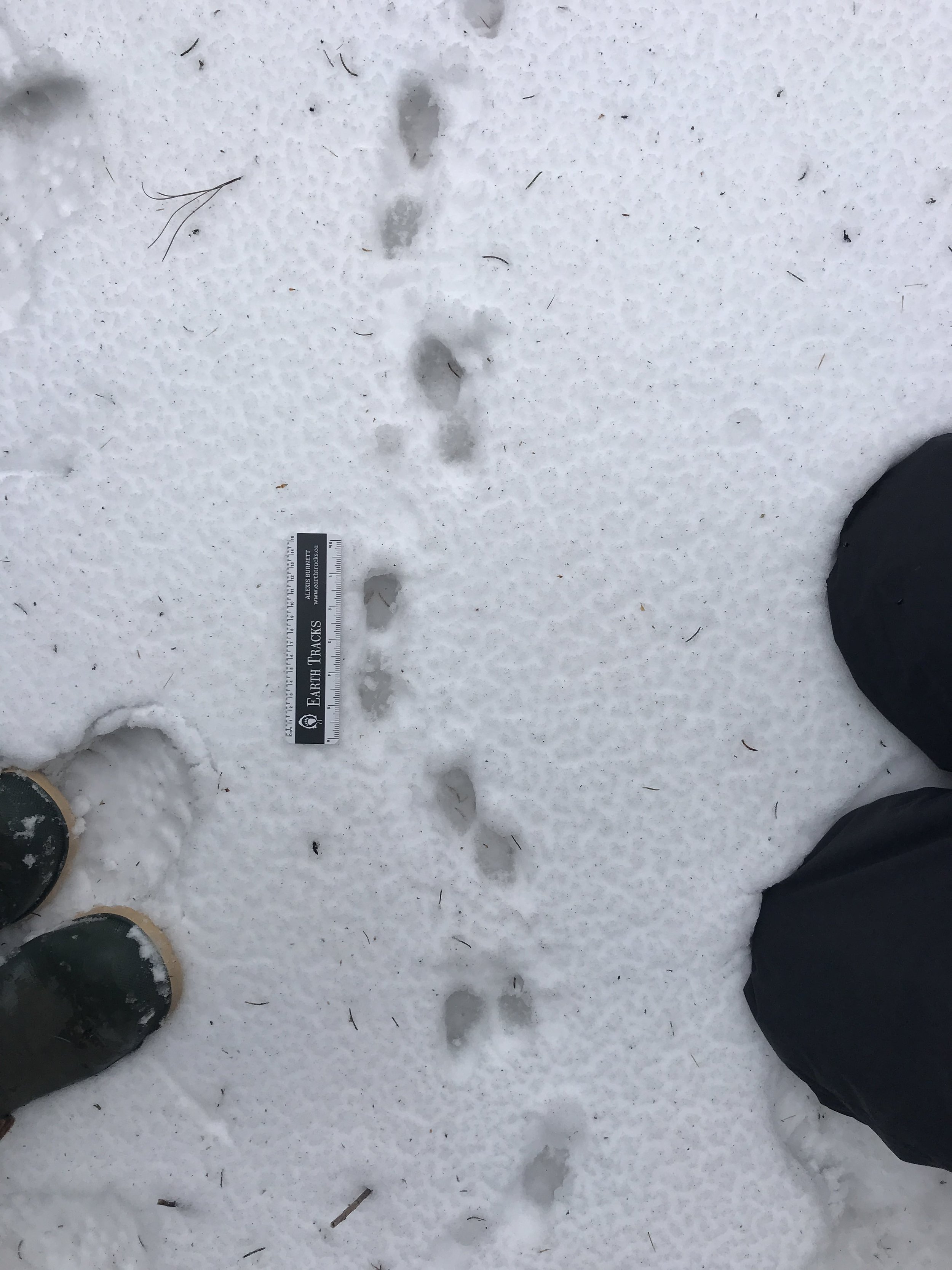
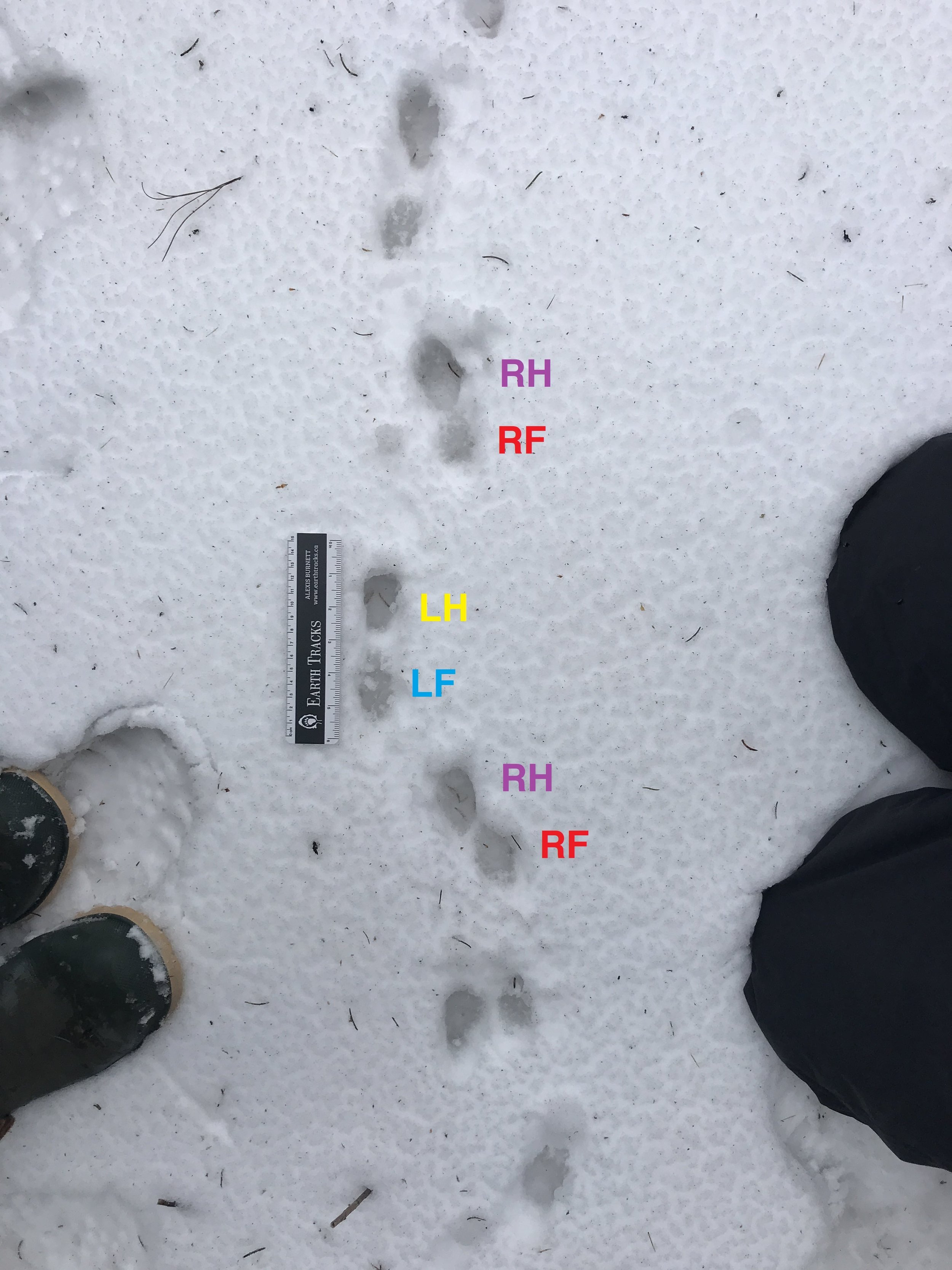
The trail shown in the fourth photo starts to show that the Skunk is slowing down. How does the trail show this? Starting with the two tracks below and to the right of the ruler, we can see that the longer track of this group of two is directly above the shorter track. The bottom of that group of two is a right front track (RF) and the track above it is from a right hind (RH). These are followed by a left front (LF) and then a left hind (LH). When examining a trail to determine the speed of the animal it is helpful to remember that the further ahead of the fronts that the hind tracks land, the faster the animal is moving. That also implies that the slower an animal is going the further back the hinds will land. Since the hinds are landing just ahead of the front tracks, we call this an overstep walk, as the hinds are overstepping the front. If the animal was moving even slower, than the hinds would land behind the fronts and we would call it an understep walk. If the Striped Skunk were moving a faster than in the photo, we would see that the right hind would land to the right of the left front track, and once these two tracks, the hind of one side of the body landing adjacent to the front of the opposite side, then we move from a walk into a lope, which is what we see in the first photo in the gallery.
Gaits can be confusing, especially when trying to explain through the internet, from someone who is still learning themselves. If you have questions, email me.
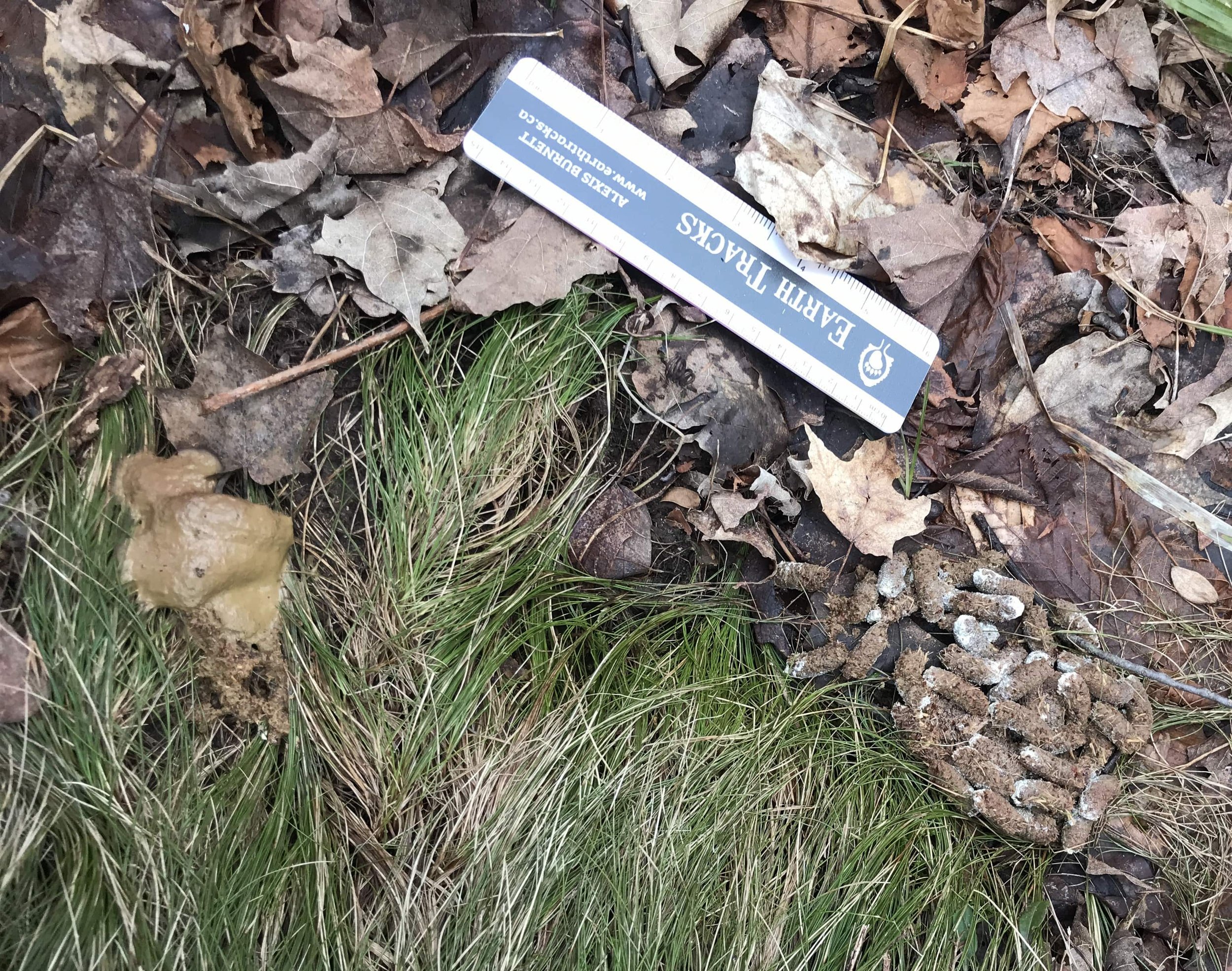
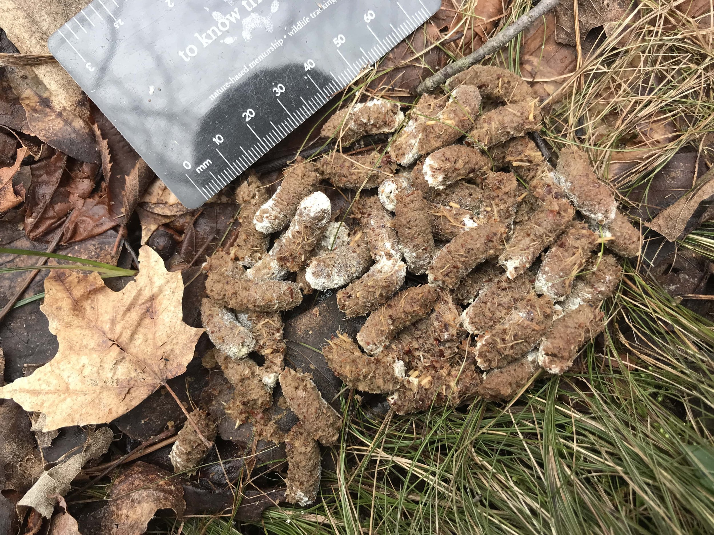
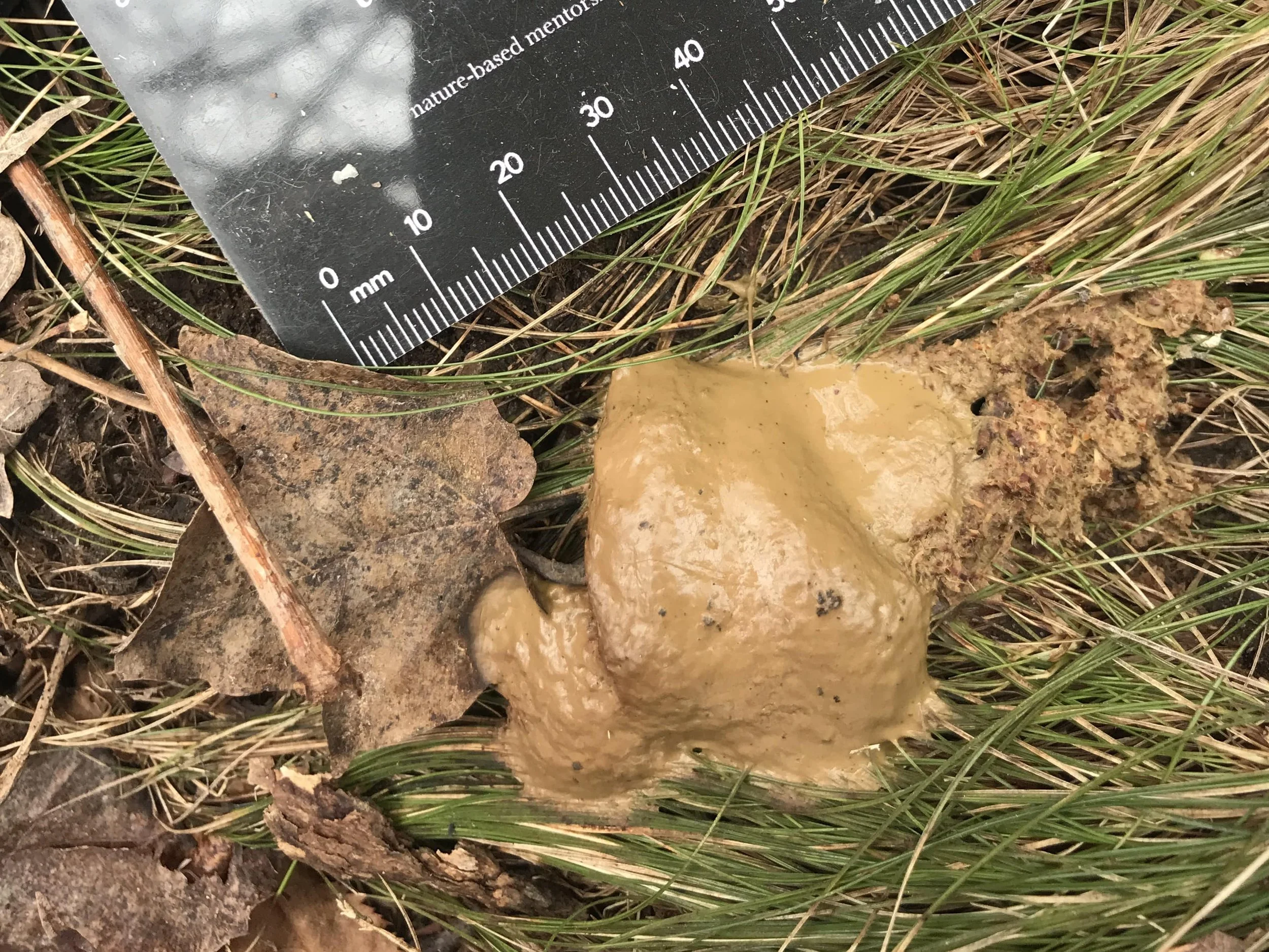
As with every outing I could go on and on as I already have, but in hopes of retaining a reader I will include one more observation. But which? Should it be a study of the strange larvae we found in the White-tailed Deer scat? What about the Oppossum (Didelphis virginiana) trail? Or maybe the old tree so decayed by white-rot that I could claw a hole right through to the hollowed out core? Instead of any of these or the many other wonders we encountered I will revisit a subject which has been irking me slightly for the past few months. Ruffed Grouse (Bonasa umbellus) cecal scat.
As I have written of before, Ruffed Grouse have two different kinds of scat. First we’ll cover their pellets. These are tubular scats about 2 cm - 3 cm (~¾ - 1⅛ in) long made up of woody materials. They are usually a pale brown with one end white due to uric acid, though I have found scats where this uric acid appears to have been washed away. The other kind of scat I find is more liquidy amorphous, lighter brown, often nearby to the pellet scat. No discernable or constant measurements as there is no constant form. This scat doesn’t last for a long time and will soon wash away as the rain comes or snow melts.
If these two different types of scat are such a constant in our tracking discoveries, what about them is confounding? Well, why are there different scats? Where in the body do they come from? Do they serve different purposes?
In Mark Elbroch and Eleanor Marks’s Bird Tracks and Sign they write
“Interestingly, after producing these lower-gut-generated solid evacuations, some game birds, such as a grouse, often then evacuate a semi-liquid brownish mass from the upper gut, or cecum, with the two types of droppings coming out sequentially; the more liquid, almost liver-colored scat comes out second and is spread on top of the solid matter. In Ruffed Grouse, it is common to find the hard, fibrous scats at one roost and the soft, brown cecal droppings at another.”
Again, I often find them together, but this isn’t the issue. The issue is complex and I might need a second to explain it.

First what the hell is the cecum? According to few sources I looked into the cecum in birds are two little pockets that stick out of the proximal colon around the junction with the small intestine. Usually they are fingerlike in shape an extend orad (towards to mouth of the bird). Some birds have big cecal pockets, but many have small or no cecum. Birds in the order Galliformes, chicken-like birds like the Ruffed Grouse, Wild Turkey (Meleagris gallopavo), Peacocks (Pavo spp.), and domestic Chicken (Gallus gallus domesticus), all have longer larger ceca compared to many other groups of birds. They also have a bit of a sphincter at the base of the ceca with small villi (tiny little projection which absorb nutrients) which may help to allow fluids to enter the ceca while other particles are passed back into the intestines through waves of involuntary muscle contractions (peristalsis).
From what I understand so far, as all materials pass from the small intestines down to the large intestine, some of the partly digested materials will be taken up by the cecum in a sort of reverse peristalsis, where it will ferment and the cellulose will be broken down further by action of bacteria which inhabit the ceca. The ceca will also reabsorb as much water as possible. The dry firm fibrous pellet scat is made up of the debris which passes through the intestines without getting to the ceca, but the wetter pudding-like scat comes from the ceca. In my research I have both read and heard folks explain that the cecal scat has a stronger scent than the pellet fibrous scat, but I have never noticed this.
However weird this muddy pudding scat seems it is actually very normal and healthy for the birds to pass, and since I have been wondering about the cecal scat of Ruffed Grouse, I believe I have since found cecal scat of Wild Turkeys, too.
Some further questions I have about the ceca and cecal scat are :
If the bacteria found in the ceca break down the cellulose, does that mean most of the pellet scat would be made up of lignin based particles?
How do parts of the intestines “know how” to separate distinct materials? Are the villi picking up on chemical constituents? Structures of materials?
Is the whole process similar with mammals who have cecum? Why do they not have two types of scat?
Above is a video I made on our outing of trying to see the consistency of the cecal scat. You can see how it’s pudding like in texture. In Bairbre OMalley’s textbook “Clinical Anatomy and Physiology of Exotic Species” she writes of birds in general who’se digestion is similar to that of Ruffed Grouse :
Some species use cecotrophy to help survival on rough forage. Food passes down the intestines to the coprodeum by peristalsis. Occasionally an unknown mechanism returns the ingesta by retroperistalsis back up to the cecum. The long villi in the cecum separate the nutrient-rich food from the indigestible portion. High nutrient particles are absorbed and the rest is passed distally in a powerful cecal contraction to re enter the cloaca as cecal droppings. These semi-solid, chocolate-brown droppings are normally discharged about once a day.
In another paper, “Gut Size, Body Weight, and Digestions of Winter Foods by Grouse and Ptarmigan” by Robert Moss he describes the digestive process as :
Briefly, the gut is generally full of digesta day and night. The crop is filled with food shortly before going to roost and this store is passed into the gut and digested during the night. After being ground in the gizzard, digesta pass through the small intestine. At the junction of the small and large intestines are two long tubular diverticula, the caeca, into which the more liquid fraction of the chyme is diverted and where it ferments. The more fibrous fraction of the chyme passes by the caecal entrances into the large intestine and is rapidly and regularly egested. The major emptying of the caeca occurs once daily when the bird is about to leave, or has just left, its roost.
Both of these quotes are better descriptions than mine above. I thought I would include them instead of trying to paraphrase. Sometimes that’s easier and just makes more sense.
—
This has been a rather long entry that has taken nearly two weeks to write. I don’t know how much sense it will make, but I find it useful to go back and review the things we know and try and take a deeper look into the how we know them, and into the literature to confirm or challenge how we know them. I want to get better at not only seeing the tracks and sign left behind, but to better understand the how and why it got left behind. From there, I want to be able to better project, to hypothesize, what other sign could look like, though imagining the forms and functions of the animals. I figure I can only do this by knowing the ecologies of the animals better, and by knowing who they are. Ironically, we can do this better by knowing their tracks and sign. I guess it’s a cycle of attention and focus.
To Learn More :
Tracking and the Art of Seeing by Paul Rezendes. Harper Perennial, 1999.
The University of Minnesota Extension page on Golden Canker
Mammal Tracks and Sign by Mark Elbroch and Casey McFarland. Stackpole Books, 2nd ed., 2019.
Medicinal Mushrooms by Christopher Hobbs. Storey Press, 2020.
Animal Skulls by Mark Elbroch. Stackpole Books, 2006.
Mammalian mycophagy: A global review of ecosystem interactions between mammals and fungi
University of Minnesota College of Veterinary Medicine website on gaits
Bird Tracks and Sign by Mark Elbroch and Eleanor Marks. Stackpole Books, 2001.
The Avian Cecum : A Review
Backyards Chickens.com article on Cecal scat vs. Pellet scat in Domestic Chickens
Clinical Anatomy and Physiology of Exotic Species by Bairbre OMalley. Elsevier Limited, 2005.
Gut Size, Body Weight, and Digestions of Winter Foods by Grouse and Ptarmigan by Robert Moss. The Cooper Ornithological Society, 1983.