
Tracking Journal 2021.10.09 pt. 2
To read the entry on part 1 of this tracking journal click here.
After most folks left, the rest of us sat down for a quick lunch. There was 4 of us left, Andrew, Rachelle, Stephanie and I. A big group can find a lot, but a small group can go at a different kind of pace, both physically and mentally. It is nice to have had the chance to have the big crew in the morning, and a smaller group for the afternoon.
Over the course of the next hour we spent a lot of time with different fungi in the Eastern White Cedar (Thuja occidentalis) forest, but I both was feeling a little bit of brain drain, and didn’t take any good photos of any of the mushrooms we were finding. I can’t quite recall who spotted the next mystery first, but it was a big unknown to me. A strange something I had never seen before or experienced before, and I have walked this trail hundreds of times. This special little treasure was handed to me and my brain clicked back to wonder and awe. What the hell was it?
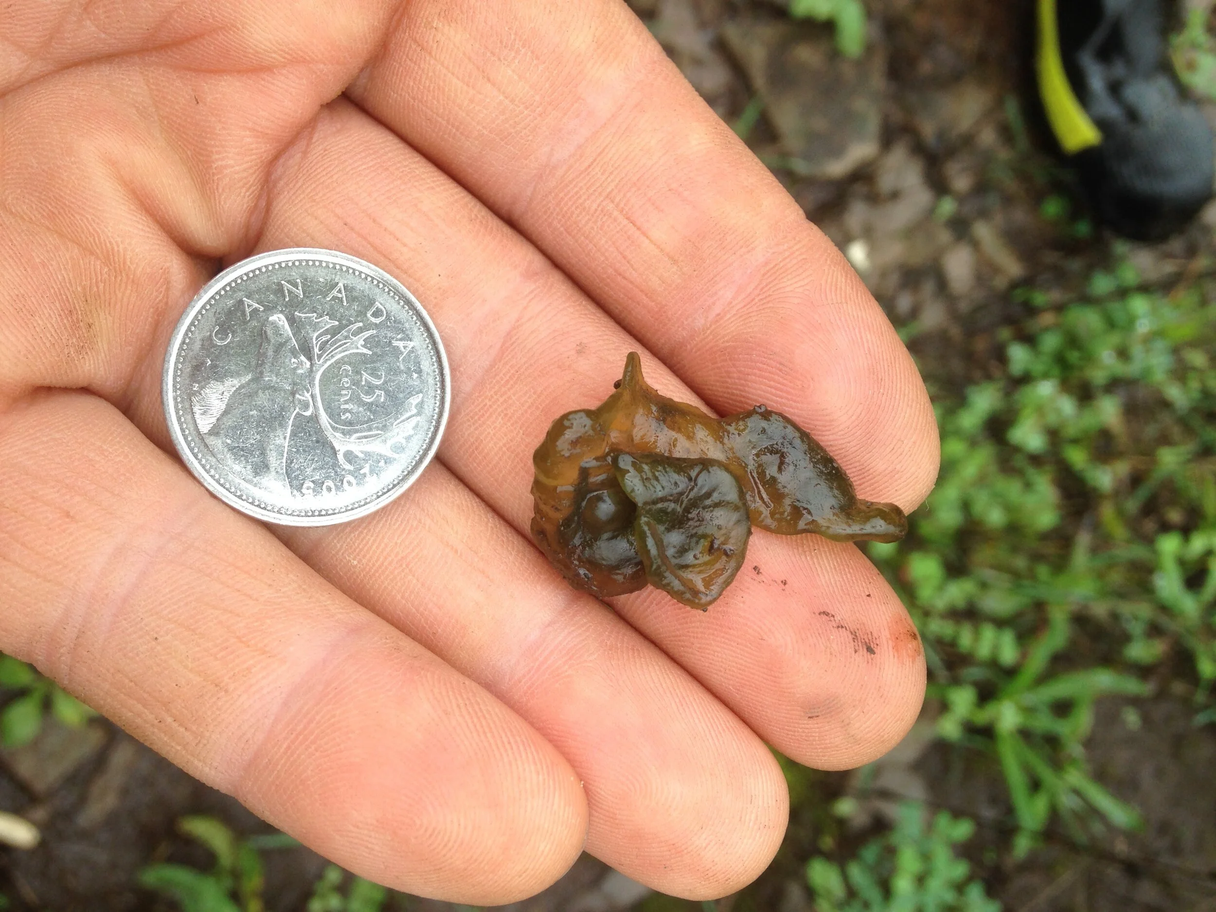
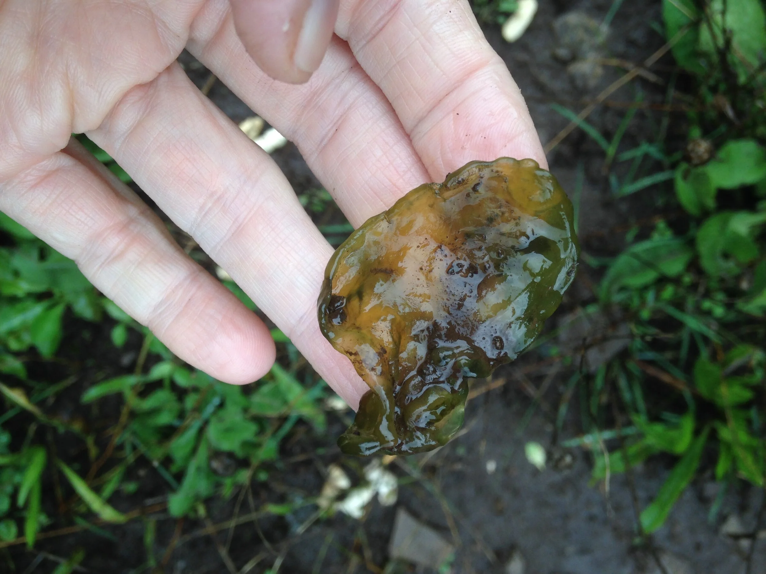
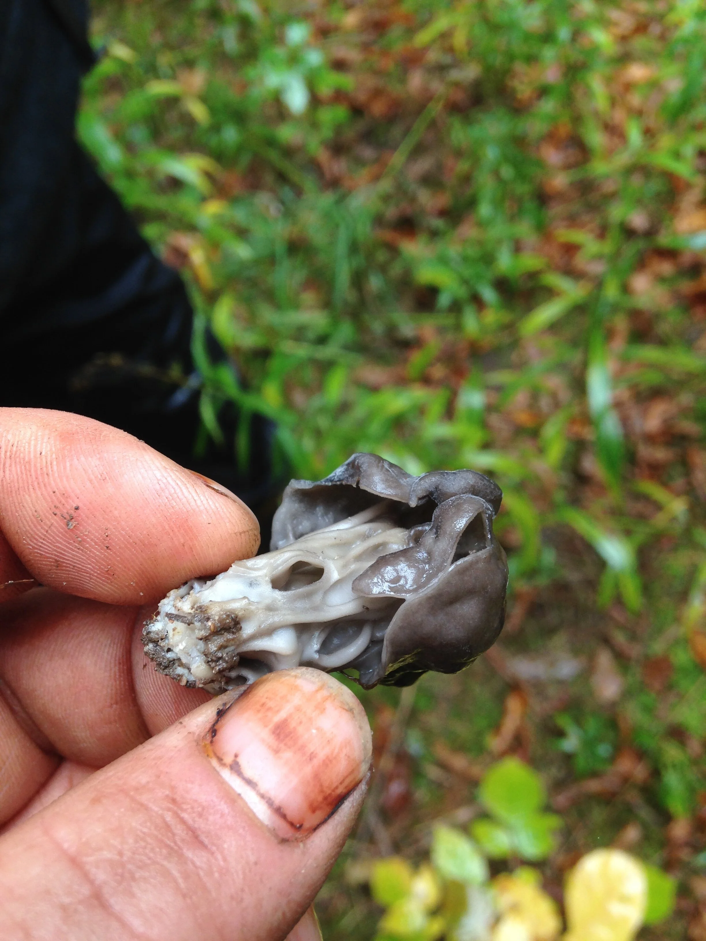
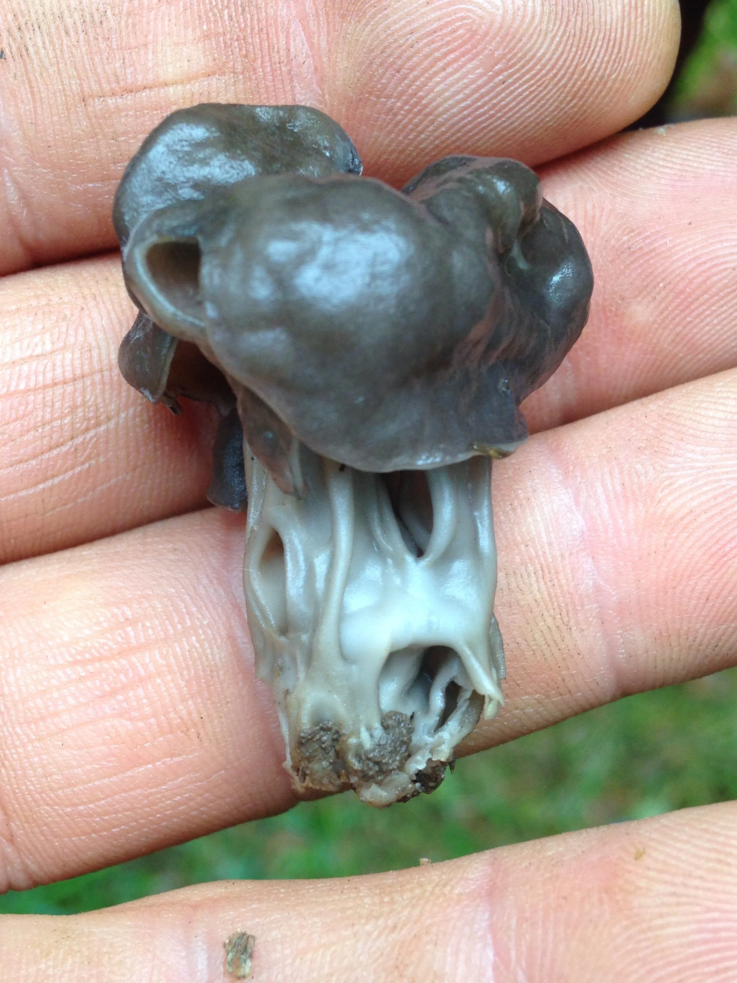
It was green and ..snotty, but a little more solid. Like someone coughed up some strange phlegmy goo and it got rained on for a while. I didn’t see it, but I think others had mentioned that there was more hidden amidst the grasses, limestone shale and small forbs. I knew I would have to look this one up when I got home and I have since looked it up. This goo has had many names, including Star Jelly, Star Shot, and Star Slime, Troll’s Butter, Witch’s Butter (not the fungi), and Witch’s Jelly. It is more formally known as Nostoc, which is the genus name of this really interesting cyanobacteria. This cyanobacteria (formerly known as “blue-green algae”) can create mucilaginous cell surfaces which allow many cells to chain together to form long filaments. The filaments can come together in gelatinous colonies built of carbohydrates with molecules consisting of a number of sugar molecules bonded together. The goo you see in the hands in the photos are these colonies!!
Each Nostoc cell contains chlorophyll, but some special cells called “heterocysts” are formed within the aforementioned filaments. The heterocysts can fix nitrogen from the atmosphere and pass it down the filaments to the smaller cyanobacteria cells who use the nitrogen to build amino acids, and proteins. The cyanobacteria in turn pass carbohydrates back to the heterocysts. Look at these cells looking out for one another.
Nostoc can live either in aquatic environments, or terrestrial environments. They can dehydrate in tougher environmental conditions, turning into a shriveled up blackish-green crust, but when the rains come, like they have been around here as of late, they rehydrate and return this slimy gooey snot body. What a lovely creation. Nostoc is probably a lot more common then what is represented by my experience on the land. It’s just I havn’t been looking, but now I will be looking everywhere.
Addendum (19.10.2021) : I have been finding Nostoc everywhere! Green little snottybodies along a trail of compacted soil I at work, especially in the puddles.
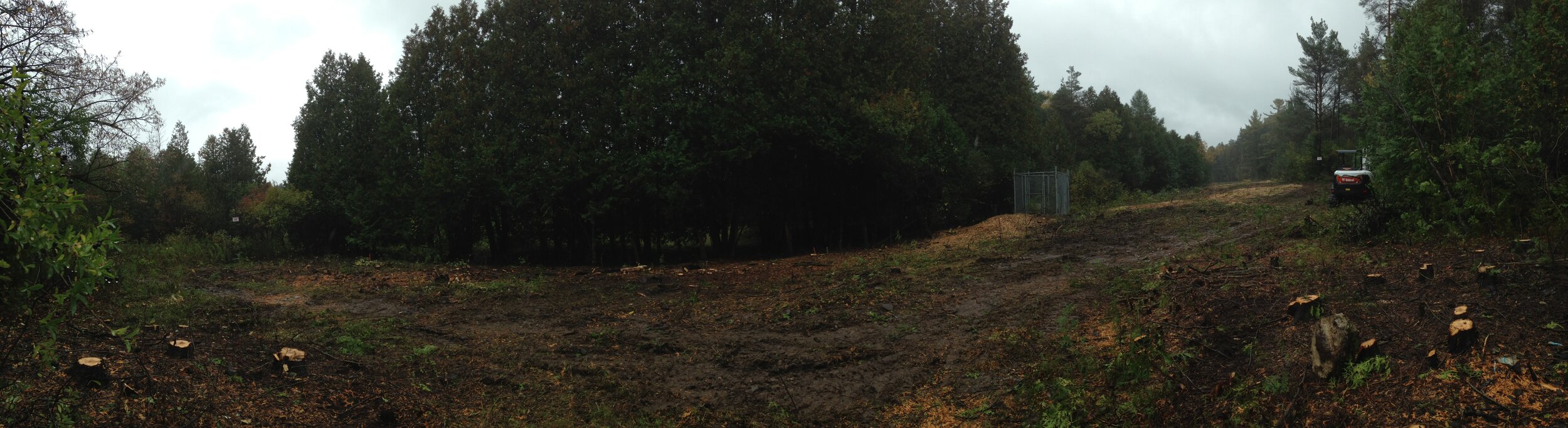
We made our way away from the Nostoc colonies and moved SouthWest towards another spot I know well. It is.. or was, a trail where I would visit a natural spring a few years ago. I would go there all the time, but lately there has been a lot of destruction going on. The city has been cutting down the Cedar forest there so they can access some watermains in the area. It really breaks my heart to see the slash where the beautiful Cedars used to be but I try and remember that the Cedar forest I had grown to love was growing atop of some previously laid watermains which were put in some 40 years before, when the watermains which had been lain before that became too rotten to work safely.
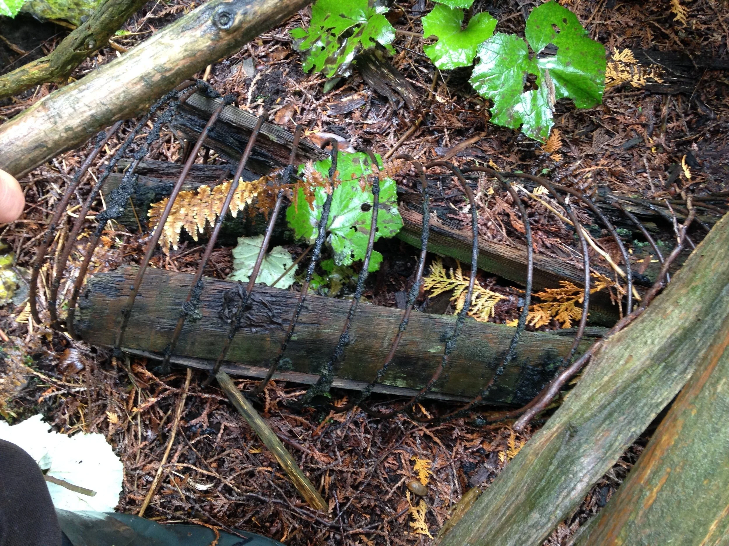
Cedar rail pipes coiled with wire.

Remnant wire coil below the Coltsfoot.
In other cities, Calgary Alberta and Hamilton Ontario come to mind, Cedar pipes were laid pre-1910 to move water from a source into the city centres. In Guelph it is the same, moving water from Arkell Springs into the city for consumption by the residents. Eventually some of these pipes began to erode despite Cedars rot resistant qualities. So the story goes that the Cedar pipes were pulled up and new tin pipes were ordered from England. On the boat trip over from the island the pipes got rattled and dented, and by the time they got to Guelph they were unusable. In light of this set back the city commissioned new pipes, this time ceramic ones.
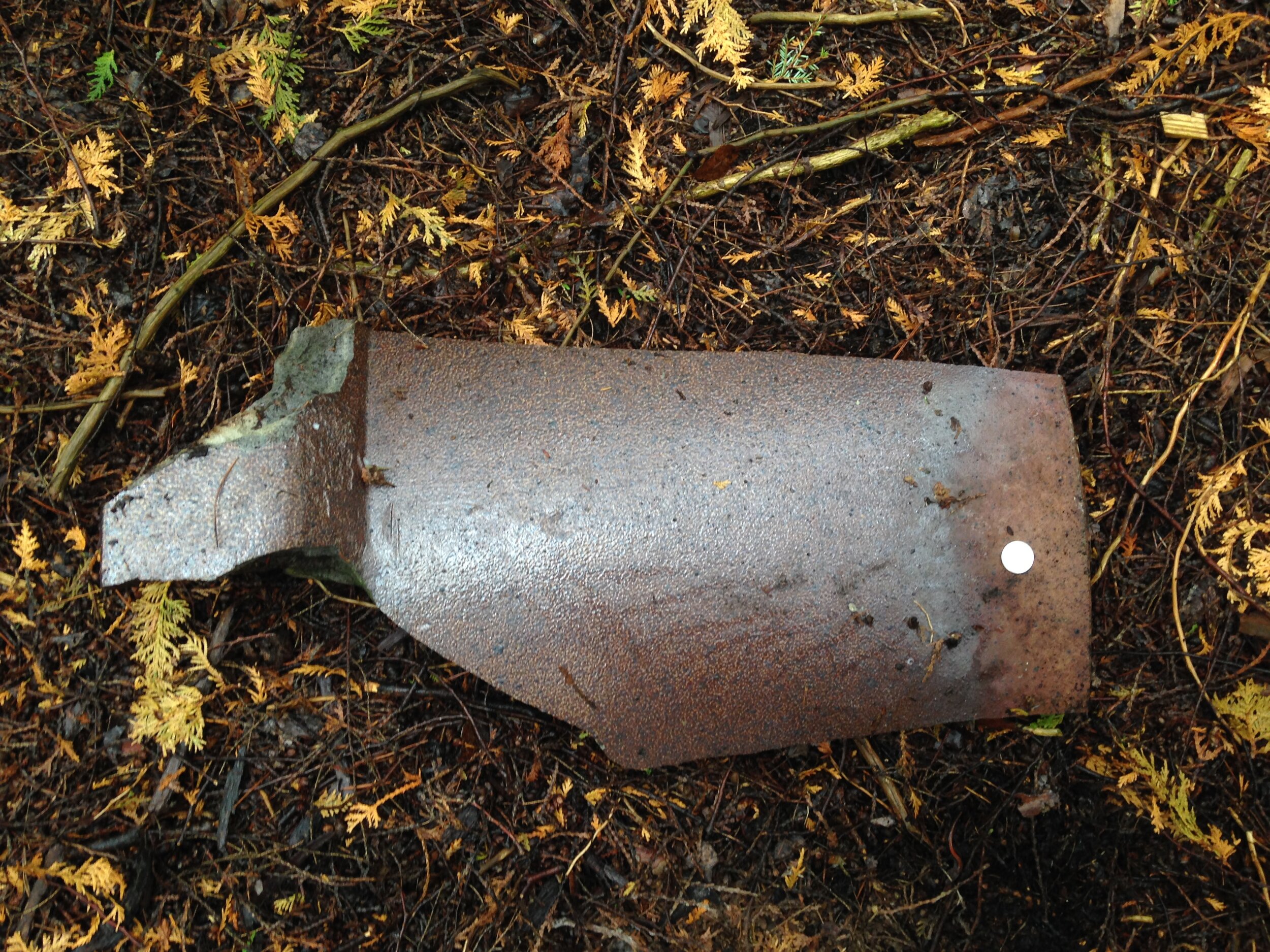
Remnants of ceramic watermain pipes with salt glaze.
Close up of the salt glaze speckling pattern.
I have seen these ceramic pipes in town recently when the city tore up my street. They are still in use in some areas and I am guessing it is these ceramic pipes which are being replaced out in the woods where they are cutting down the trees. The intention is to keep the landscrape cleared this time by building a gravel road atop the pipes allowing for easier vehicle and mechanical access to the pipes if and when they need to be repaired or replaced. As my friend Arlene shared with me today, I recognize the need to cut the forest down intellectually, but it still hurts the heart to see all those trees get cut down.

We continued on through the now light drizzling rain in search of a planted White Pine (Pinus strobus) plantation where I had been a few days before. We were still on our search for more mushrooms. As we stepped into the shade of some tall Spruces (Picea spp.) which bordered the Pines everyone got excited about how many mushrooms littered the ground. Not just the amount even, but the variety. So many mushrooms that you had to watch every step you took lest you might step on some. The Pestle-shaped Puffballs (Handkea excipuliformis) were ripening, and the thrill of poking them was just too much. I ended up picking up one of the ones pictured (the one on the right and squished it repeatedly while watching the millions of spores float off behind me. I couldn’t stop squishing it. Some deep joy filled me as I did so and reminded myself of the chance that will come when I get to smash up some of the Giant Puffballs (Calvatia gigantea) later in the fall.
But something else caught my eye. Down on the ground, just below a small White Ash (Fraxinus americana) was a tiny little purplish grey mushroom with a strange cap and even stranger stalk (stipe). I didn’t know this mushroom but it looked a little like a Morel gone funny. Perhaps one of those False Morels (Gyromitra esculenta)? But someone else knew better. Someone called it an Elfin Saddle.

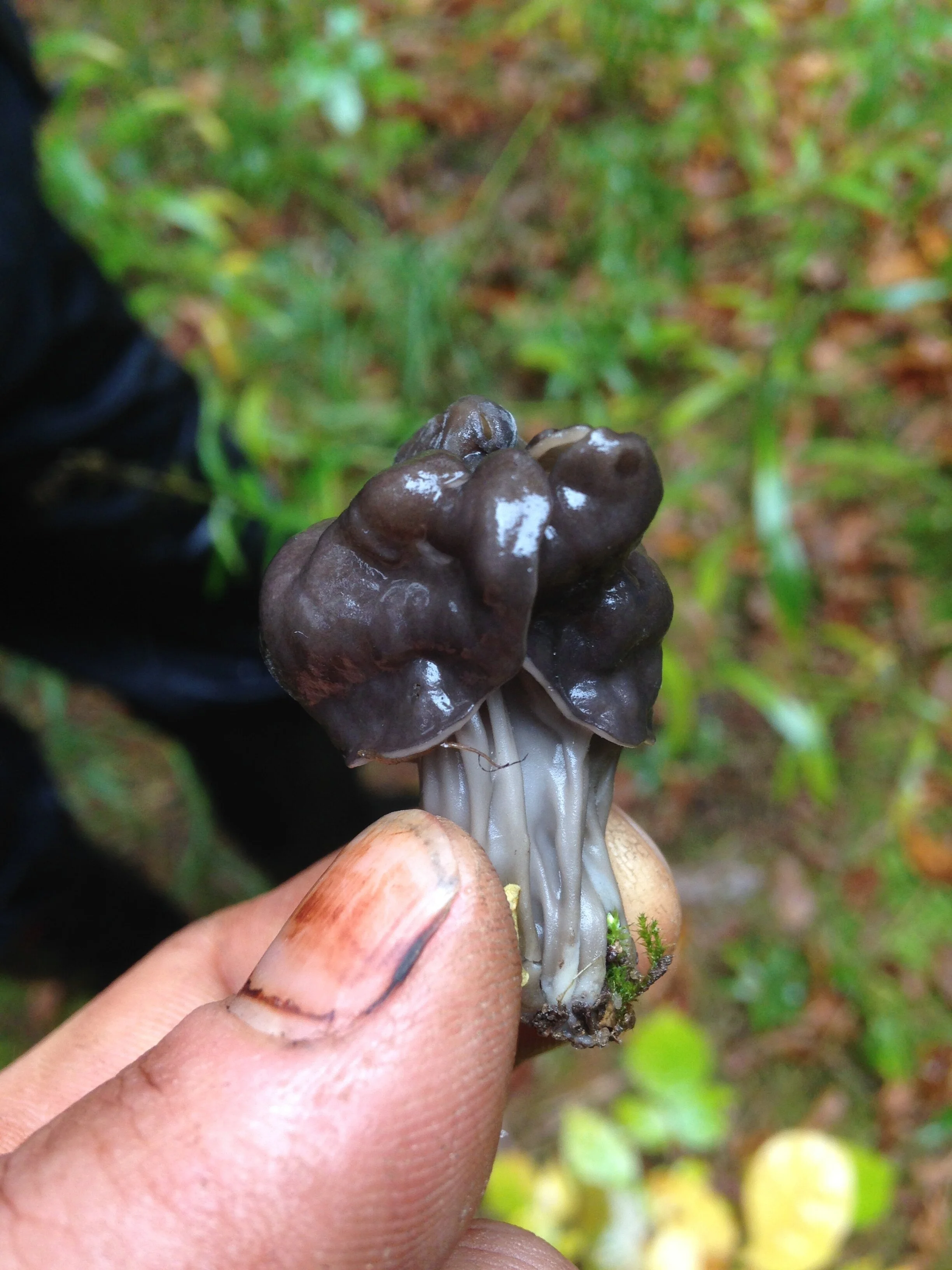
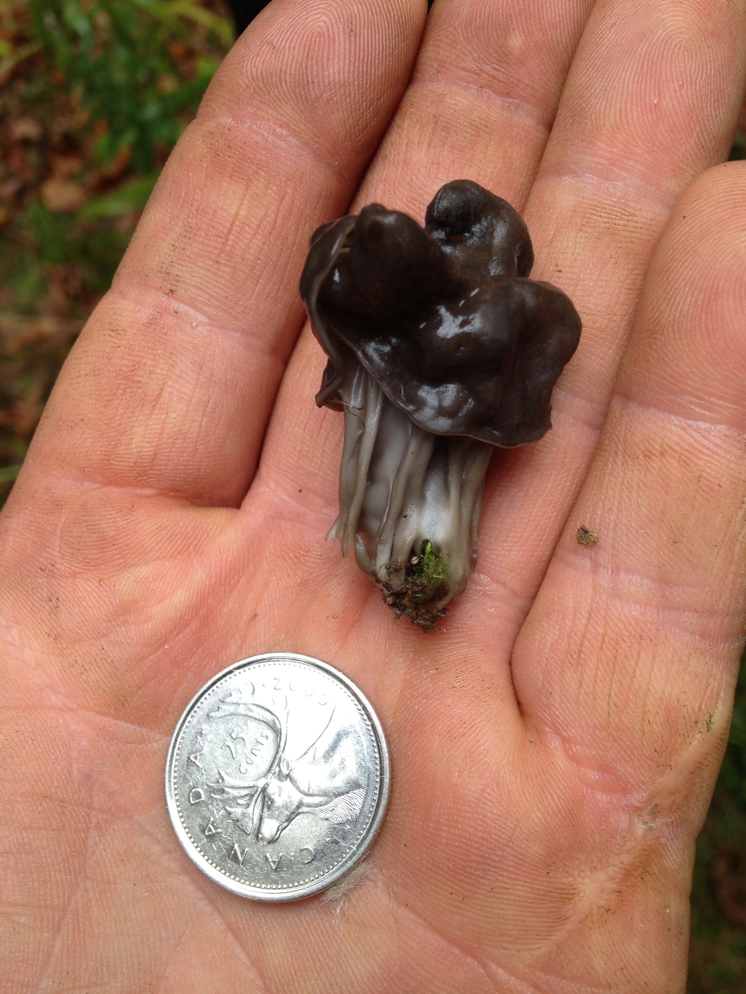
Turns out they were right. It is indeed an Elfin Saddle. It is Helvella lacunosa, known as the Fluted Black Elfin Saddle. Both names, I decided, would make for a good black metal band. I was really intrigued by the stalk and who might make their home there. Were there any known arthropod associates with the H. lacunosa? This really seemed like the kind of mushroom that fairy tales would include. I have seen Amanita’s growing close to this spot in the Summer, but now I think this may be the most bad assed mushroom on the block.
One book I have describes Elfin Saddles as “grow from the ground near living deciduous trees an conifer; they may also grow in grassy locations, along paths and in disturbed areas.” Andrew brought up the question of sample bias in regards to where we might find these mushrooms. Of course we will see them growing often along paths and disturbed areas, as those are the places that humans commonly are - inevitably we will encounter more of everything there. What Andrews question brings up is another one, of where else do/would the Fluted Black Elfin Saddle grow that humans may not spend as time time or interact with? Do they grow in the thickest of nasty smelly swamps full of dangerous stuff? Or high inaccessible cliff walls? Unlikely, but yeah, where else may H. lacunosa spend their time? It was an interesting question for me and something I think I will be considering into the future.
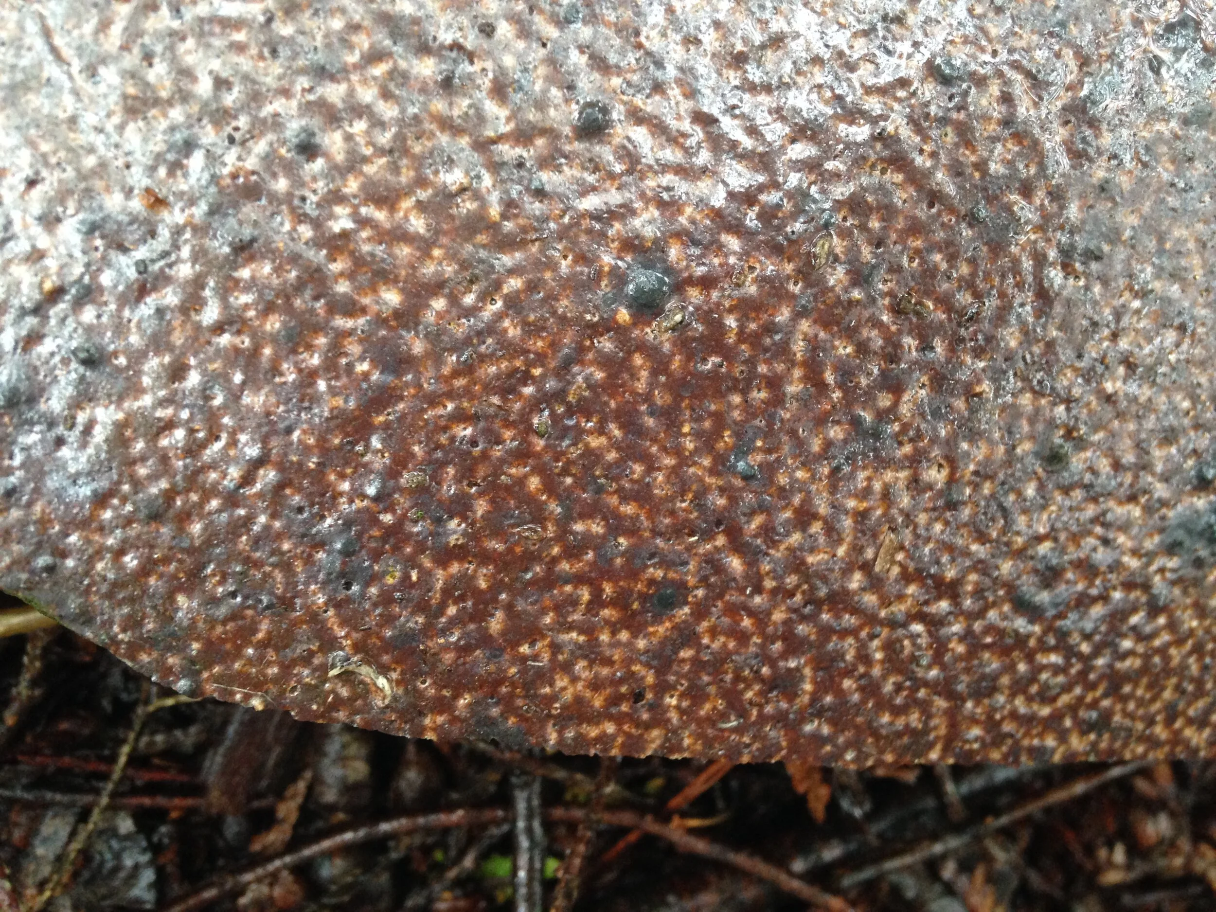
In the same plantation we noticed a growth on a couple of the Pines which I just couldn’t ignore. It was so prolific on a couple of the trees, but most of them were seemingly untouched. This strange fungi? looks like a similar something affecting some Eastern White Cedars (Thuja occidentalis) at work near by.
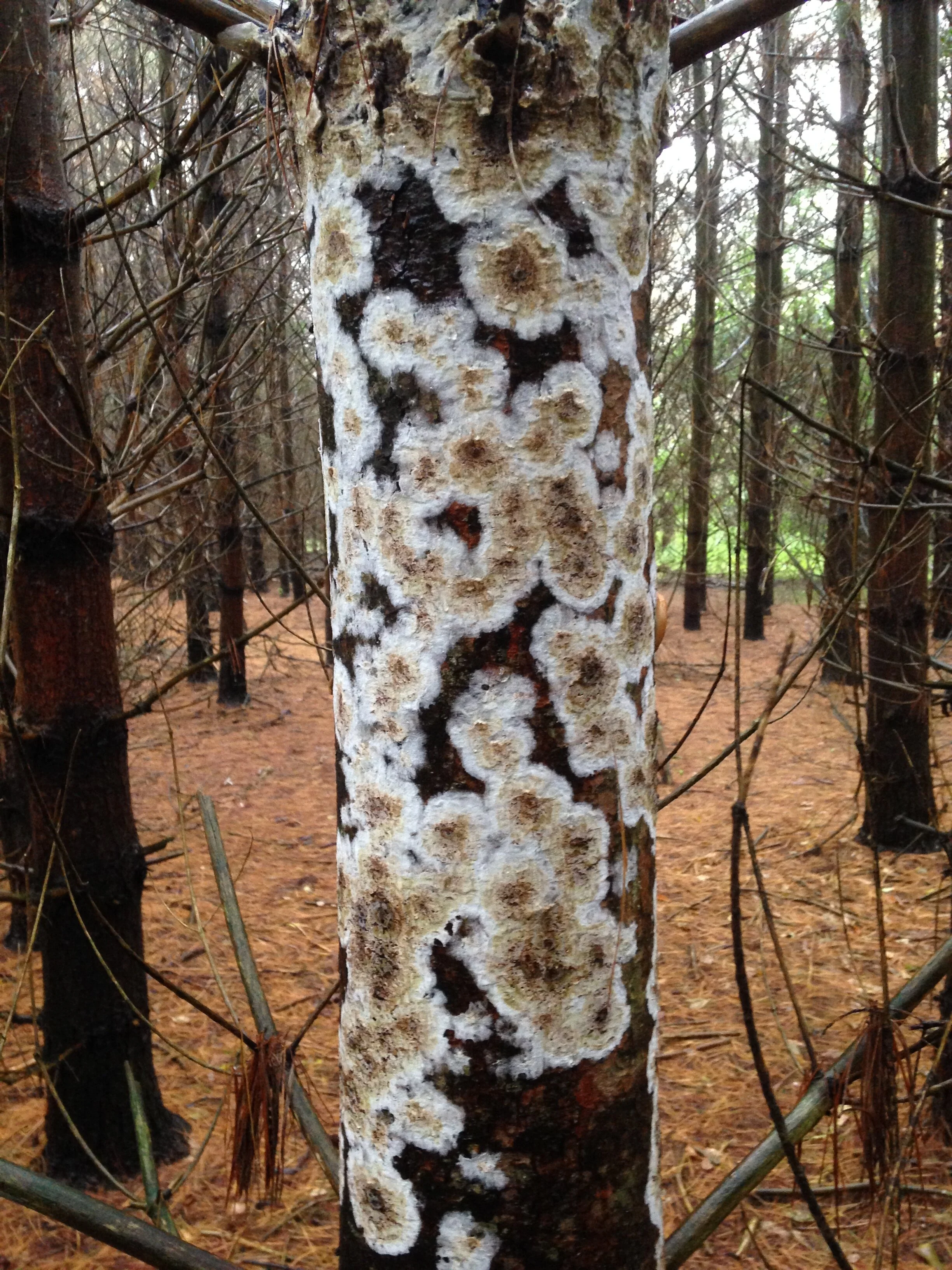
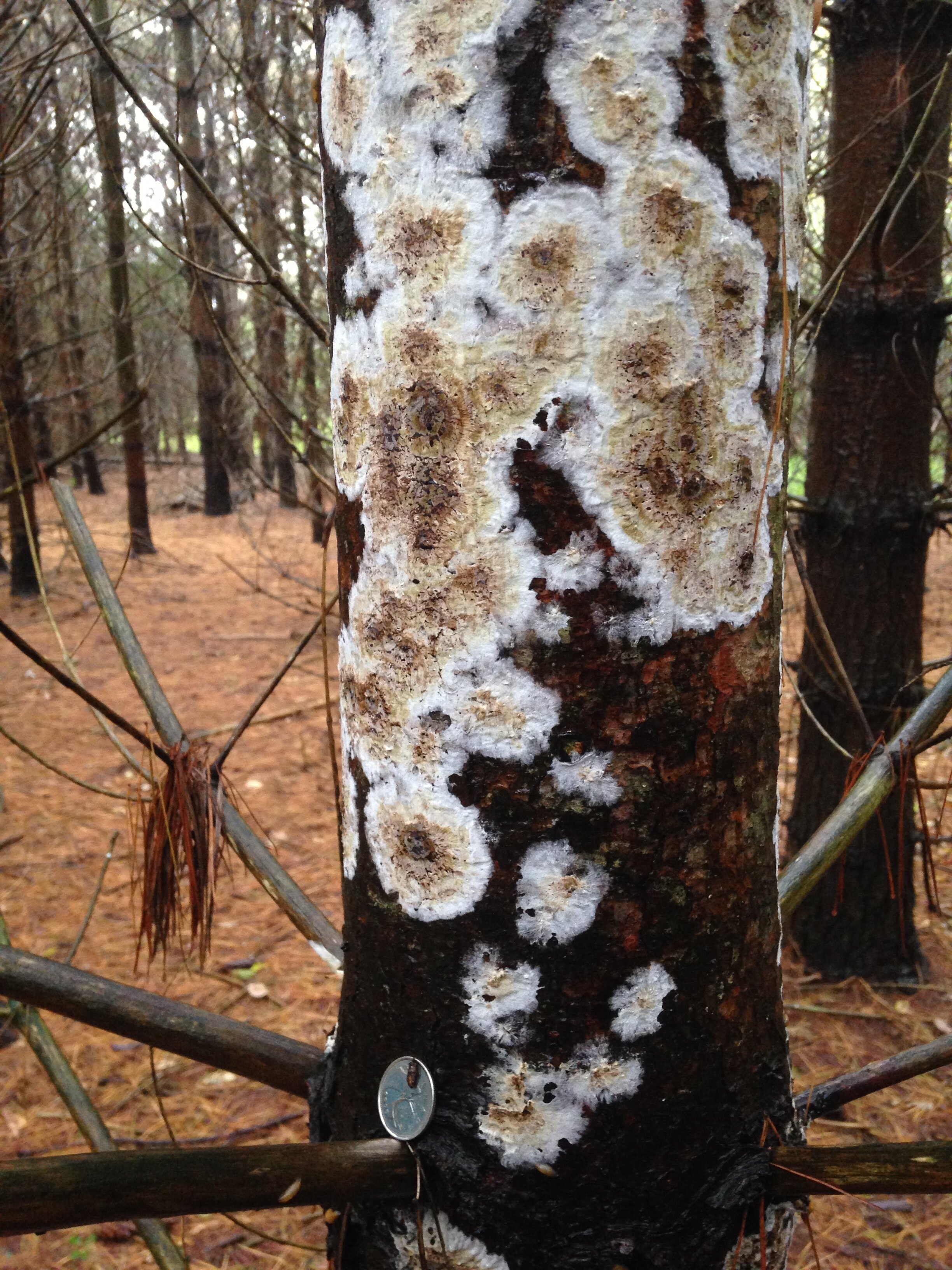
Above is the White Pine, and below is the White Cedar.
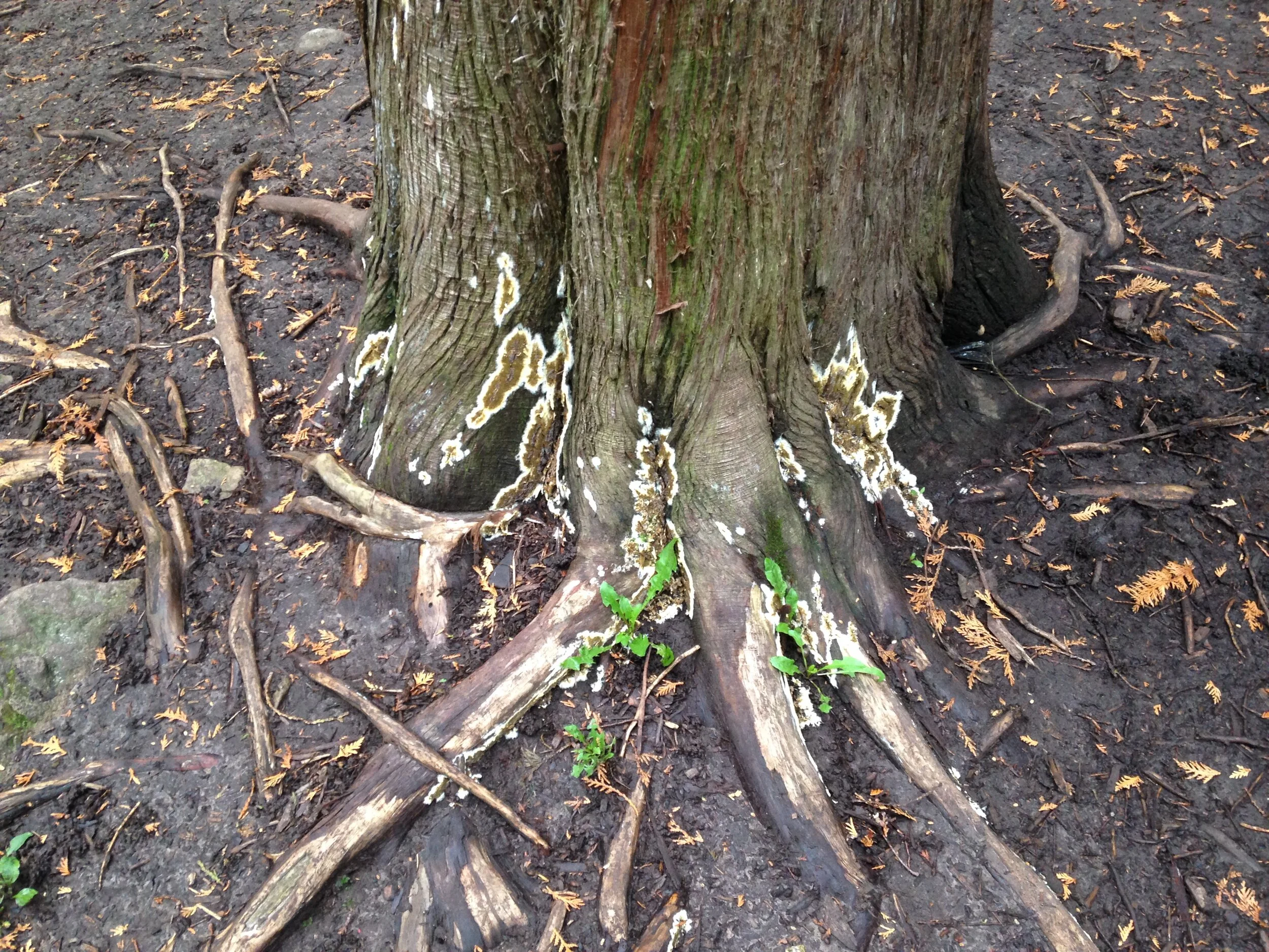
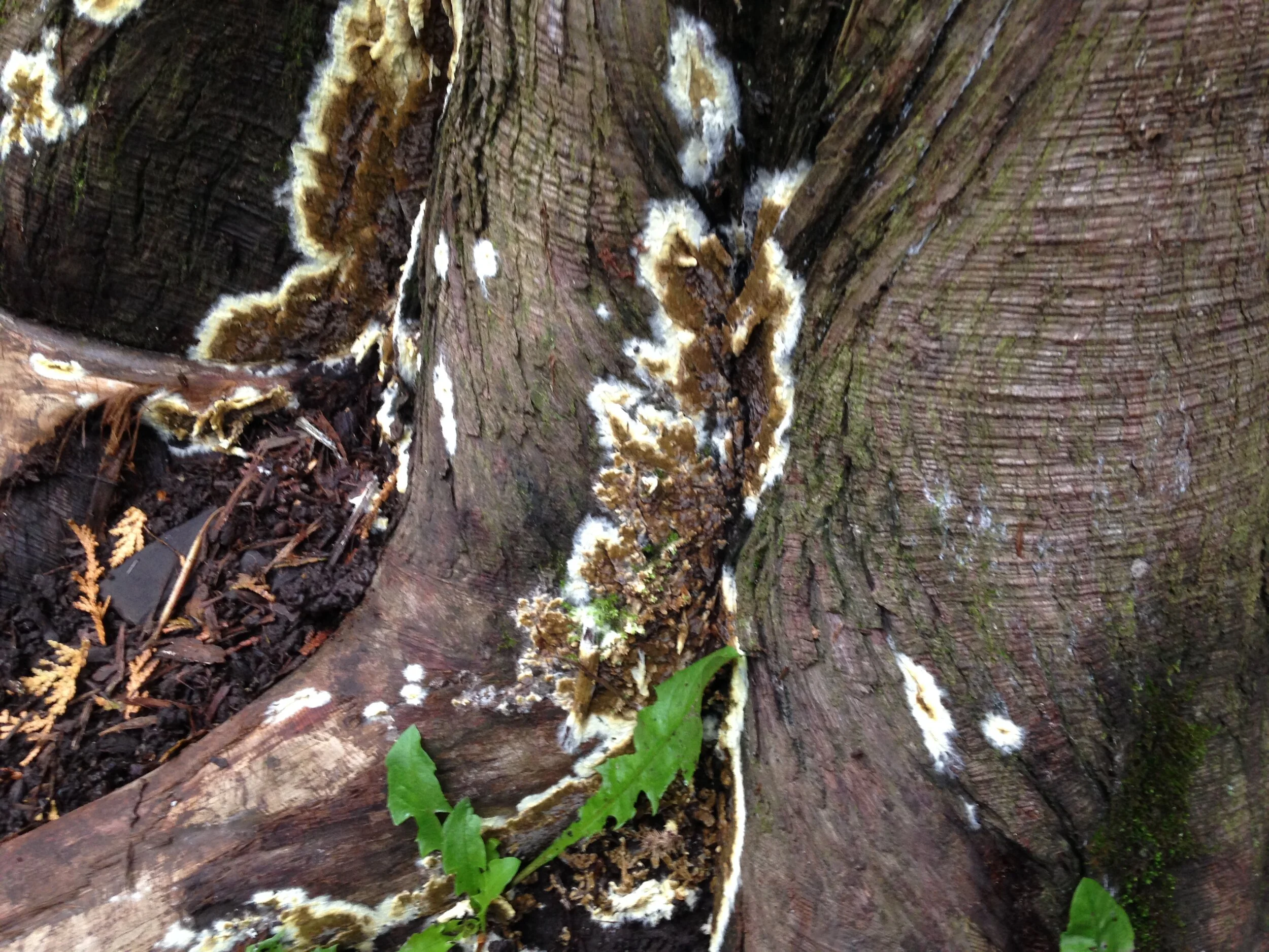
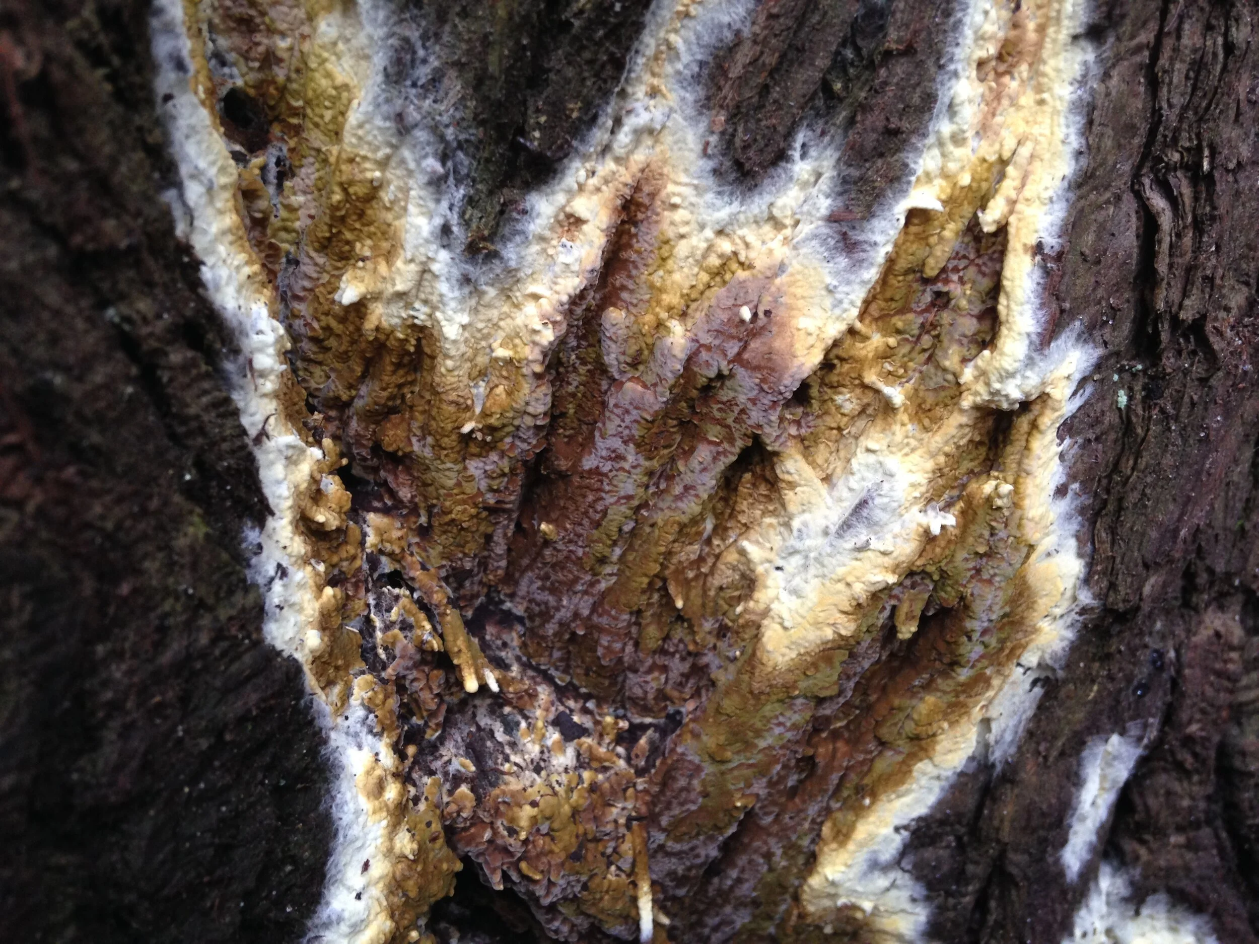
After some research, including asking on the internet, and google image searching I believe that this species of fungi is Olive Duster (Coniophora olivacea). I am still not 100% but it seems to be close enough to call it. The closest case of Coniophora olivacea I found on inaturalist.org was in Kirtland, Ohio, which isn’t too far - it would be like driving from Guelph to Tobermory and back, so a fungal species with that sort of proximity seems likely, even if not reported North of the lake.
Addendum (19.10.2021) : A pal who was at the first half of the outing, Matt, suggested that this could be another species in the Coniophora genus, Coniophora puteana or Wet Rot. When comparing this species in inatualist, it has come up more often in Ontario. I am still looking for information on how to differentiate the two Coniophora species, but until then I want to keep this open.
From what I have learned so far, Coniophora are basidiomycete fungi belonging to the order Boletales that produces brown-rot decay on dead wood of conifers. Brown-rot fungi, produce oxalic acid an hydrogen peroxide which break down cellulose fibers in smaller sections which are eventually broken down even more into sugars that the fungi gradually consumes. There are different kinds of brown-rot, one of which is called “butt-rot” which I thought was hilarious. But really, butt-rot just means rot associated with the trunk of a tree. So, it could be explained that this is an olive coloured butt rot fungus, or perhaps Olive Butt Duster could be a new common name?

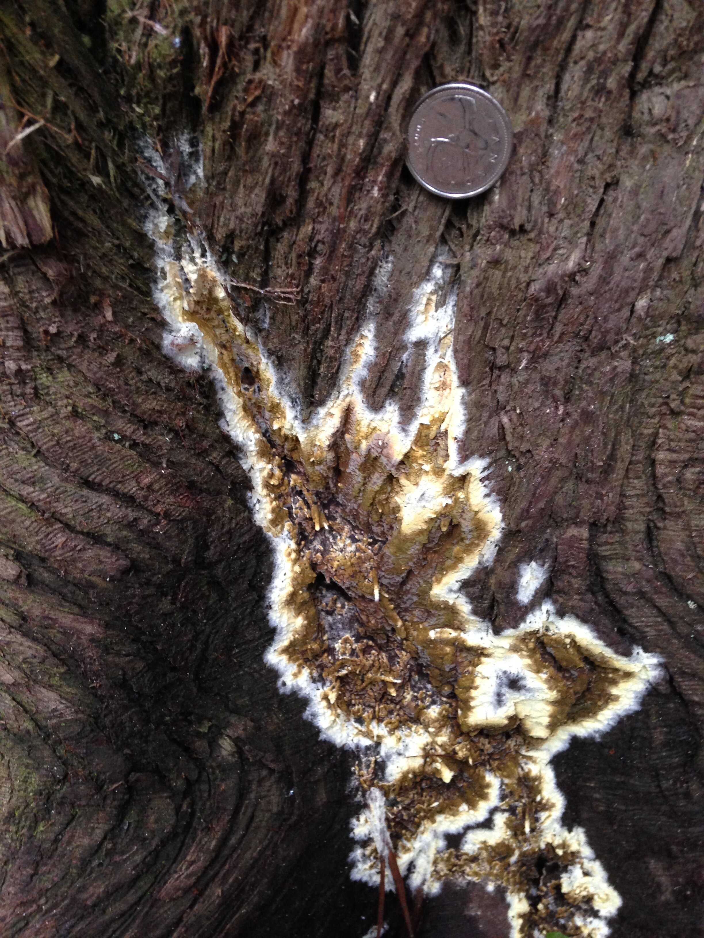
An example of brown-rot which I photographed a week after the tracking club outing, 15.10.2021
White rot is created by the process of fungi digesting cellulose and the extremely durable lignin parts of the wood and demonstrates by leaving behind stringy fibrous strandlike pieces of pale cellulose. White rot breaks things down a little more thus freeing up more nutrients trapped in the wood. Brown-rot is created by the fungi digesting the cellulose, but leaving the lignin. It is demonstrated, as in the image, by leaving cubeish residual lignin. I have seen brown-rot breaking down to finer and smaller cubist pieces so that eventually it crumbles easily and becomes the humus soils found in some forests. Noting the pattern of rot can help as an extra sign in the process of i.d.’ing different fungi. The decaying wood of a single tree can be caused by both white-rot and brown-rot at the same time.
Anyhow, back to tracking, or at least wandering around the woods looking at cool stuff..
We wandered slowly through the Pine plantation, still checking out all sorts of mushrooms, some of which I had previously photographed and written about, and some of which I was glossing over. I think brain fog can be a thing for me and likely for other trackers out there. When I practice paying attention and trying to note a lot of what is happening around me, from the movements of birds up in the canopy, to faraway sounds, to what my companions are up to and talking about, to considering the trail we should take… it’s like pilates of the the mind. It gets tiring after a while, and I become disinterested for short stretches. I do really think though it is an exercise and can be made stronger with more practice, and variability. Perhaps this is when I can start to play some games to switch the parts of my body and brain are working the hardest, and give myself a little moment of chill. Maybe a sit spot, or a fast running game, something to bring me back to a focused awareness and refreshing my spirit. I think I was also getting hungry which may have also been a component to my brain fog so I suggested that we start heading back and we made our way a little bit quicker through the plantation out towards an opening in the forest which was mostly dominated by a creek fed pond. Grasses and other vegetation had been taking over the muddy edges of the pond for a few years, encroaching on the waterline and stabilizing the soil. We watched a couple of Flycatchers on a huge dead Cottonwood (Populus spp.), and took a seat on the busted up length of another Cottonwood which had fallen a couple of years before. This change of scenery, and quick sit was enough to get me up and wandering around again.
The last time I had been in this spot I had been watching Argiope aurantia, or Yellow Garden Spiders, sitting in their webs, amazed by their size, colour, and overall appearance. They were truly beautiful. But when I showed the photos to colleagues from work, one co-worker said he had just found some of their egg sacs! He described finding them in Goldenrod flowers, at about belly button height (we are generally the same height, ~173 cm, or 5’8”). He found his in the dark with a flashlight, so I figured I might as well look if I had all the daylight I wanted, and knew that the spiders were around. So I went and looked. In the tall grasses closer to the remnants of the pond I noticed a small something amidst the Goldenrod and grasses, but it turned out to be a Goldenrod Ball Gall, and not an egg sac. I was a little discouraged, and was telling Andrew about it. He left to go explore around the fallen Cottonwood while I kept looking. I had seen photographs of the egg sacs recently in a book somewhere so I had the image in my mind which I was comparing everything I was seeing to that specific search image, when I looked deep into the tall grasses in front of me and saw one.
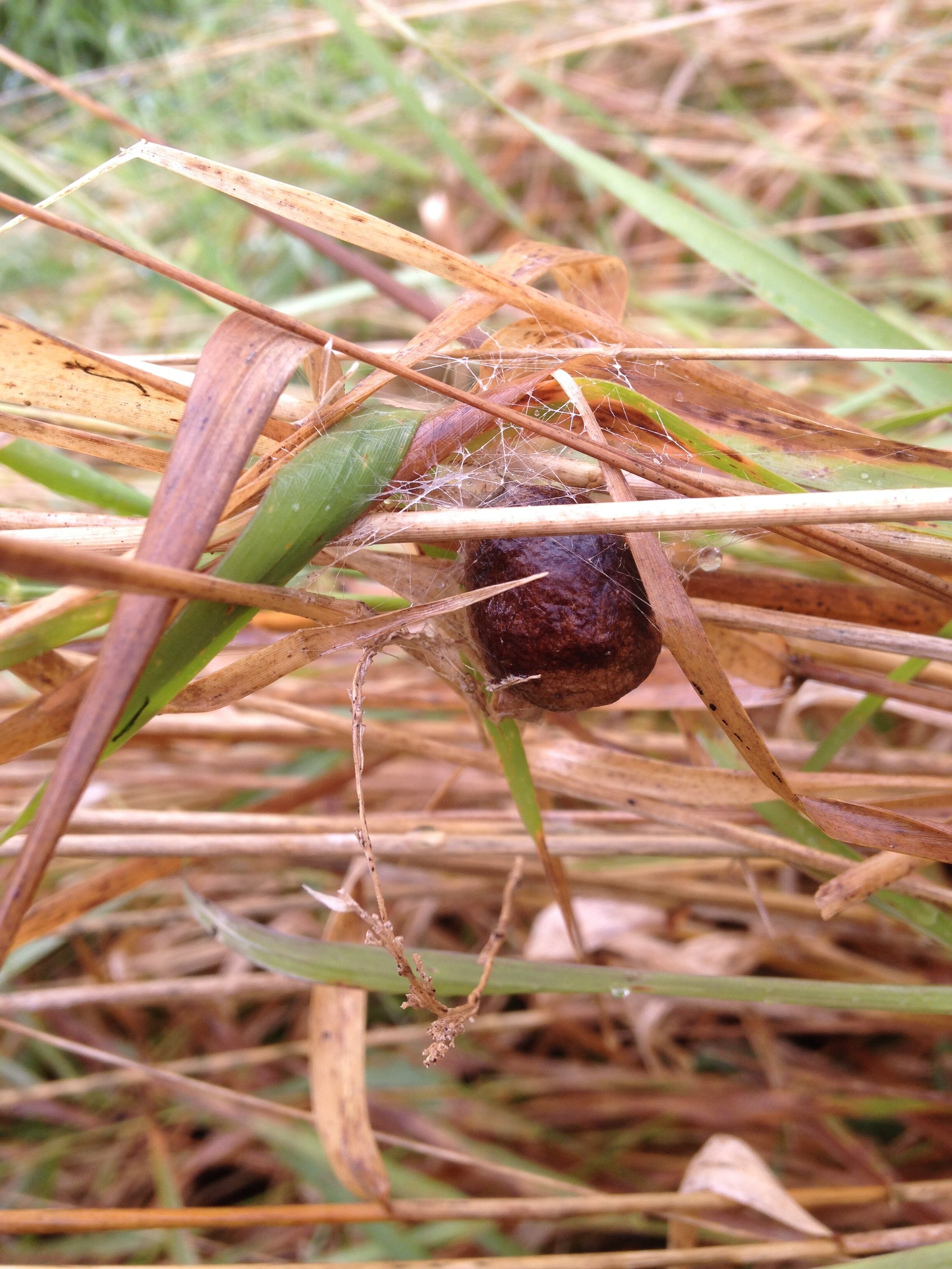

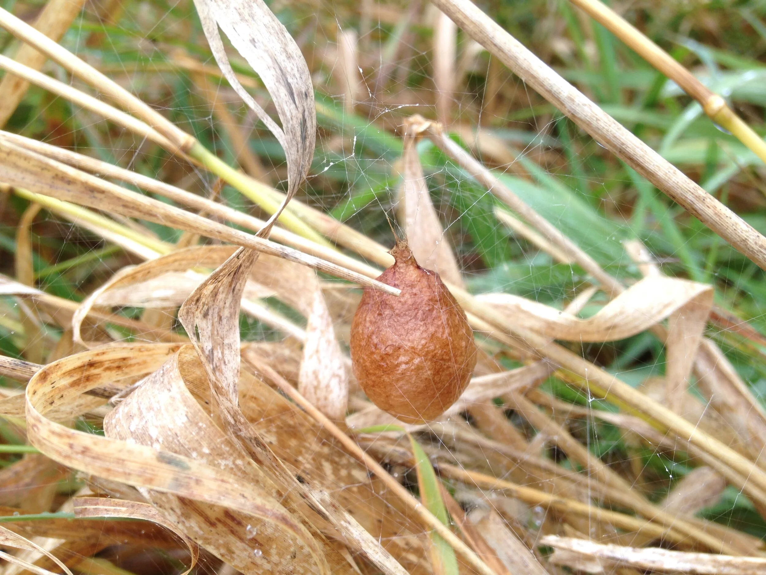
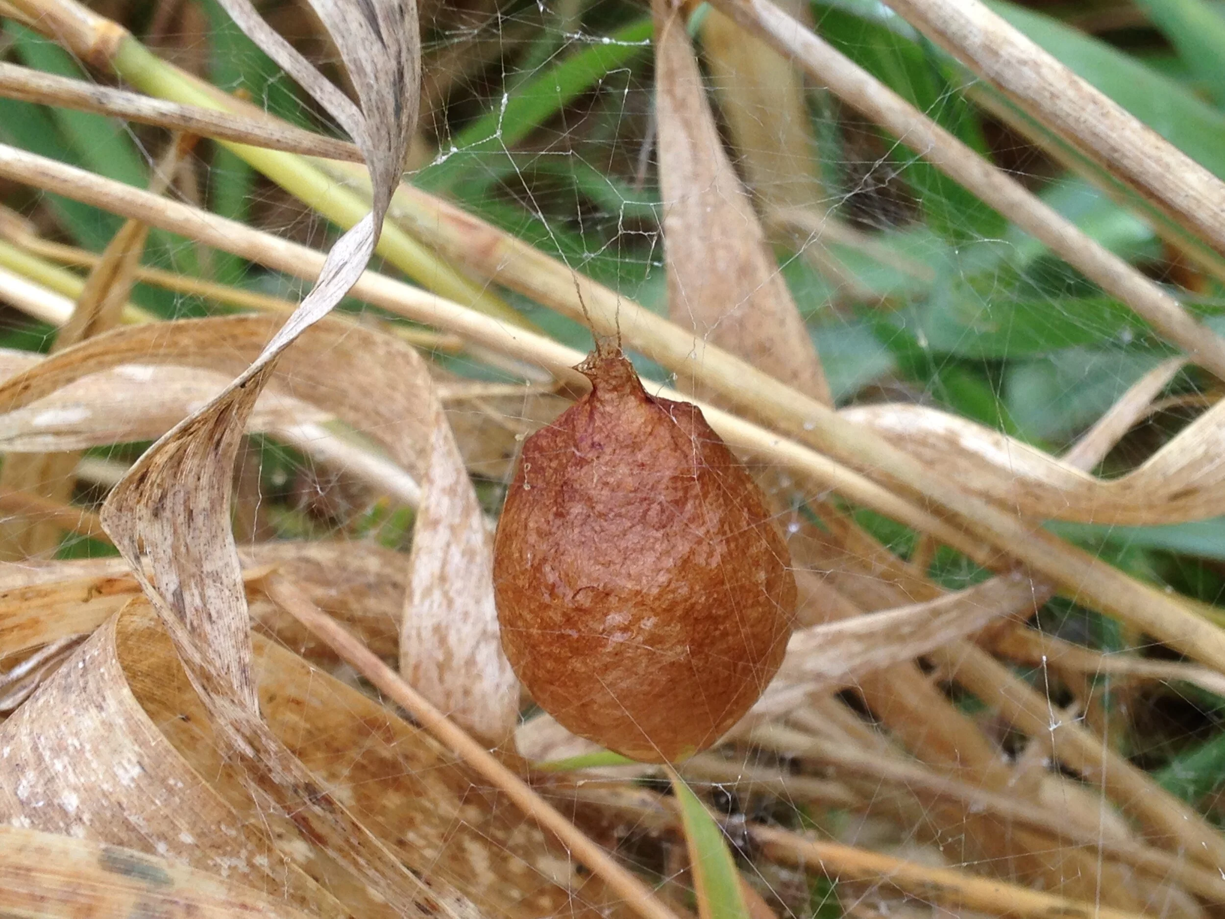
I saw one, and then shortly afterwards I saw another. I was elated! These egg cases are pretty magical, not just for A. aurantia, but for a bunch of different species. But we’ll get there. There is so much I am learning, and so much more I’d love to know.
The egg sacs in the images above a little older, but not by much. When they are first created they are whiteish to yellowish and turn the tan papery brown in time. How much time? I don’t know but it would be cool to understand the aging process so you can tell how long since the egg sac had been created. The egg sacs in the images typical of Yellow Garden Spiders that I have seen in books and online. Often found in long grasses, Goldenrods (Soliago spp.) and other taller forbs in the Fall. They can be 2.5 cm (1”) long, spherical, with a narrowing at the top. There are actually three different layers of silk which make up the matts and eventually the casing for the eggs. If we were to stay up late, head down to the pond at night to watch the female build these egg sacs, she would first be creating a soft silken layer on which the eggs would be placed. Next, she would cover that matt with a fluffy layer of silk which I would guess acts as a bit of an insulator against the cold, and then finally the outer papery tough layer. I imagine the papery outer casing may be similar in feel and durability as a Cecropia Moth (Hyalophora cecropia) cocoon which is extremely durable and resists tearing. Ultimately, I don’t know for sure as I didn’t touch the egg sac (not out of ick factor, instead I didn’t want to disturb it too much). Spiderlings hatch in the late Fall but overwinter inside the egg case, only to emerge in April or May by collectively chewing a hole and through enzymatic digestion (the process of breaking down material into easily absorbed and assimilated substances by the action of enzymes) in the tougher strands in the outer shell of the egg case in the Spring. I have included a linked image (below) from the study where I learned this.

Image from “Relation between the outer cover of the egg case of Argiope aurantia (Araneae: Araneidae) and the emergence of its spiderlings”, by Foradori et al, 2002.
I have read in another paper I was looking at that the mean (average) number of spiderlings which emerged from the 114 egg cases collected was 341. One source, which I sadly cannot remember, wrote that the eggs will hatch at different times, and the larger, earlier hatched spiderlings may predate on their younger siblings.
All the research I was doing around these egg sacs got me thinking more deeply about how I access knowledge, share that knowledge and how I credit the knowledge I have.. brought up a lot of questions, which for the time being are still questions, but something I would like to write about sometime in the future.
There were a couple more highlights for me from the tracking outing last Saturday; the Sweat Bee (Agapostemon spp.) we found on the Cottonwood, or the Gummosis on the Black Cherry (Prunus serotina) tree, or even the Ruffed Grouse (Bonasa umbellus) wing and feet Rachelle shared back at the parking lot. But there was one that really stood out as we were walking back to the cars along the train tracks, someone noticed another dead snake.
I can’t remember who it was that had found the snake, but it was dead for sure. Mouth was frozen wide open, with sign of visible trauma just behind the head - the snake was flattened in that area, and a break about 3 cm above the tail. I don’t know what killed the snake, but I knew it was dead. I remember picking up the snake and looking at them closer. Another Dekay’s Brown Snake (Storeria dekayi), which made for two total for the few hours we had been out. I moved the snake a little on my hand and Stephanie asked if the snake was still alive. I figured she saw the snake move when I moved my finger, so I did it again and I think she, and maybe the rest of us, noted that the snake had not moved on their own, but only because I moved the snake… until the snake moved again. I hadn’t moved my finger this time, but just behind the flattened portion, maybe a quarter of the way down from the head, something inside the snake began to move. Of course this was met with squeals of joy and disgust. I set the snake down on the railroad ties and Rachelle volunteered to begin the dissection.
It did take sometime after I got home to confirm that it was not a wasp larva. I had been wondering about the parasitizing of caterpillars by wasps for a couple of weeks, and so they were on my mind and I thought this must be a case of that exact thing happening.. but the larva that crawled? scurried? wriggled away did not look like any sort of wasp larva that I had seen in the images or videos I had been watching. I had to go home and search online through different images of larva, maggots and even an inchworm or two. I got down to two families within the Diptera (Flies) order. I am thinking either Blow Fly (Calliphoridae family) or Flesh Fly (Sarcophagidae) larva, and cannot yet determine which is more likely the one we found. Their method of wriggling about, the wider back end and narrower head, the size, the timing, and the broader context, I really do think it is one of these larva but I cannot determine between them. I even found a key to some European species that may help to tell the larva of the two families apart, but I would need a microscope and a few specimens. We didn’t bring that larva home, but watched them disperse. One cool distinction is that the Blow Flys lay their eggs in the carrion (oviposition), and the Flesh Flies lay their larva (larviposition). Very fun.
Blow Flys have earned a new respect in my legendarium since researching for this entry. I have been reading about how they can smell a decomposing organisms on wind currents from up to 2km!!! away, and locate the animal within minutes of dying. They are very timely decomposers. Forensic entomologists, folks who study the insects associated with a human corpse in an effort to determine elapsed time since death, among other related data, try to understand the relationships of Blow Flies and the dead things, especially humans which they inhabit. Through understanding the stages of life of a Blue Fly, they can determine how long someone has been dead for by looking at how mature, or developed, the Blow Fly, or other insects, are. That’s totally rad. That’s tracking wildlife in a whole other level.

© 2012 Nature Education Adapted from Tomberlin et al. 2011. Used with permission.
Later on, I got to worrying about the maggot themselves. Was it appropriate to push them out of the snake’s body just to learn something? In follow up research I have learned that the maggots will often leave a host, burrow into the soil and enter into their pupal stage there in the soil, and not in the previous host. This dispersal is a normal part of the Blowfly life cycle. We may have hurried the process, but the maggot should be fine baring predation from birds, other inverts or even another snake.
We made our way back to the cars where Rachelle shared he Ruffed Grouse parts and pieces with us, and were we finished our day off with another round of gratitude and sharing any questions we were taking with us. I know I didn’t answer everything I was wondering about, but I did get around to some of them.
To learn more, check out these links:
Teaching the Fungal Tree of Life
Spiders of Toronto (pdf)
Blowflies Help Determine Time Of Death
Forensic Entomology: The Use of Insects in Death Investigations